
Author: Dermatological Society — Updated 2025
What is angiolipoma?
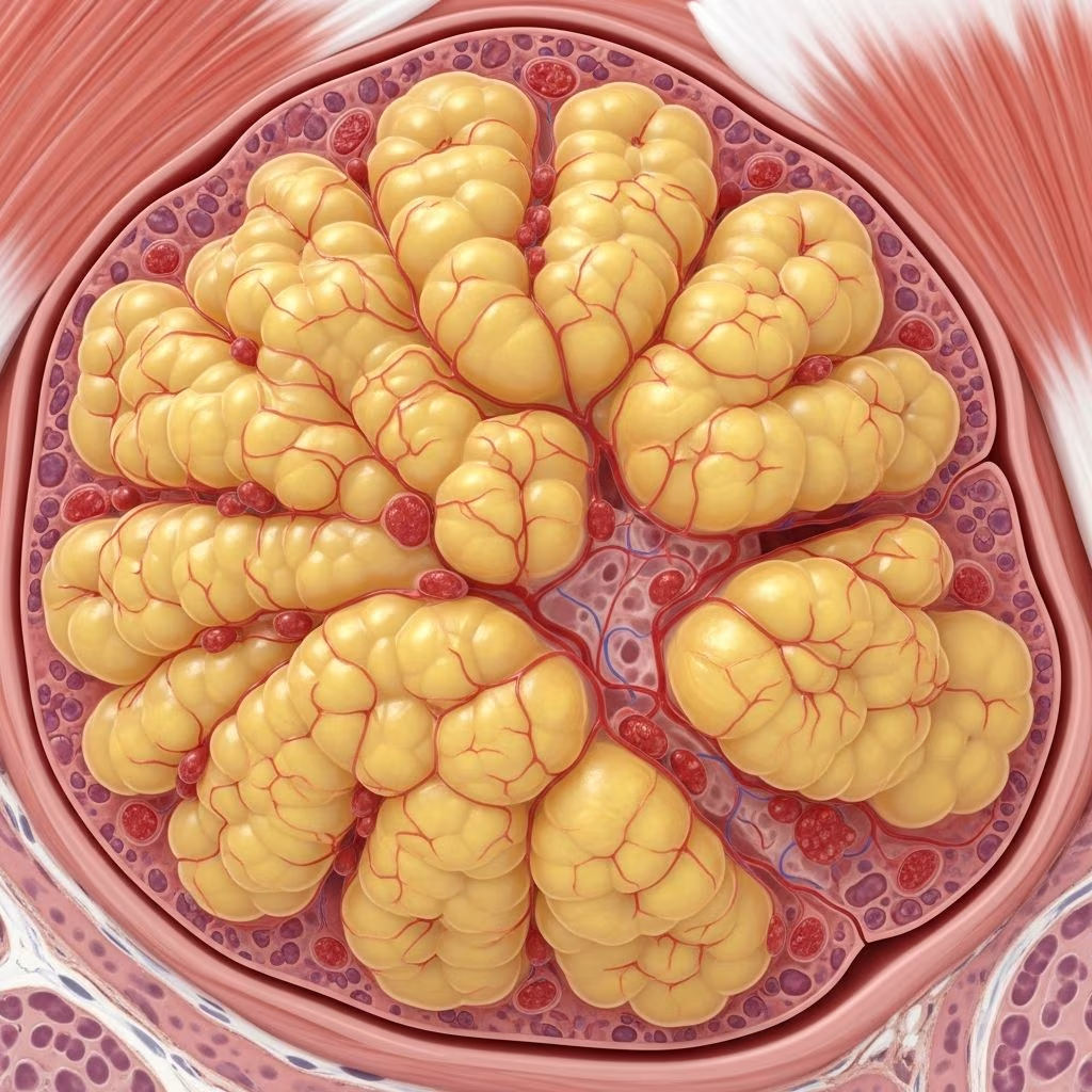
Angiolipoma is a benign subcutaneous tumour composed of mature adipocytes (fat cells) and a prominent vascular component of capillary-sized blood vessels. These lesions are classified as a subtype of lipoma but distinguished by their vascular proliferation and frequent pain on palpation.
Angiolipomas typically present as multiple, tender, subcutaneous nodules, most commonly on the forearms of young adults. They account for 5-17% of all lipomas and occur 3 times more frequently in males. Unlike typical lipomas, angiolipomas are often painful due to their rich vascular supply and presence of fibrin thrombi within capillaries.
The term ‘angiolipoma’ reflects its dual composition: ‘angio’ referring to blood vessels and ‘lipoma’ to fat tumour. They are considered hamartomatous rather than true neoplasms, as no consistent genetic abnormalities have been identified.
Who gets angiolipoma?
Angiolipomas most commonly affect young adults aged 20-30 years, with peak incidence in the second and third decades of life. They demonstrate a 3:1 male predominance.
Lesions often arise post-puberty, though the reason for this age predilection remains unclear. Familial cases with autosomal dominant inheritance have been reported, suggesting genetic predisposition in some patients. Trauma has also been implicated as a precipitating factor in occasional cases.
Clinical features of angiolipoma
Angiolipomas classically present as multiple painful subcutaneous nodules, though solitary lesions occur in approximately one-third of cases. The forearm accounts for ~65% of sites, followed by upper arms, trunk, and lower limbs. Less common locations include head/neck and hands.
Key clinical characteristics include:
- Size: Usually 1-4 cm diameter, rarely exceeding 4 cm
- Shape: Round/discoid, well-circumscribed
- Consistency: Soft, doughy, rubbery, or spongy
- Mobility: Freely mobile over underlying structures
- Skin: Normal overlying skin without discoloration
- Pain: Tender to palpation (hallmark feature distinguishing from typical lipomas)
Two clinicopathological subtypes exist:
- Non-infiltrating angiolipoma (90%): Well-circumscribed, superficial subcutaneous
- Infiltrating angiolipoma (10%): Extends into muscle/fascia, higher recurrence rate

Diagnosis of angiolipoma
Diagnosis relies primarily on histopathological examination, as imaging lacks specificity. Clinical suspicion arises from painful subcutaneous nodules in characteristic distribution of young adults.
Imaging features
- Ultrasound: Hyperechoic mass with prominent vascularity on Doppler
- CT: Hypodense fatty mass ± enhancing vascular septa
- MRI: T1 hyperintense (fat signal) with T2 hyperintense vascular components
These modalities demonstrate the fatty/vascular composition but cannot reliably distinguish angiolipoma from other fatty tumours.
Histopathology
The definitive diagnosis requires biopsy/excision showing:
- Mature adipocytes forming the bulk of the lesion
- Peripheral capillary proliferation forming small vessel clusters
- Fibrin thrombi within capillaries (diagnostic hallmark, present in ~100% cases)
Two histological patterns exist:
| Pattern | Fat:Vascular Ratio | Features |
|---|---|---|
| Classical | Predominantly fatty | Peripheral capillary aggregates ± thrombi |
| Cellular | Equal or vascular predominant | Numerous capillaries throughout, few adipocytes |

Differential diagnosis
Key differentials include:
| Diagnosis | Distinguishing Features |
|---|---|
| Lipoma | Painless, minimal vascularity |
| Leiomyoma | Firm, smooth muscle bundles |
| Neurofibroma | Nerve sheath elements, S100+ |
| Haemangioma | No mature fat component |
| Liposarcoma | Atypical lipoblasts, pleomorphism |
Treatment of angiolipoma
Asymptomatic lesions require no treatment. Symptomatic lesions are managed surgically:
- Non-infiltrating: Simple excision, <5% recurrence
- Infiltrating: Wide local excision with margins, 35-50% recurrence risk
Surgery performed under local anaesthesia. Postoperative pain relief often dramatic.
Alternatives: Steroid injections may shrink lesions but less effective than excision. Liposuction suitable for superficial non-infiltrating lesions.
Investigations
Clinical examination sufficient for most cases. Imaging (US/MRI) if deep/suspicious. Excisional biopsy provides diagnosis and treatment.
Possible complications
- Surgical site infection (<1%)
- Recurrence (infiltrating subtype)
- Cosmetic deformity (large forehead lesions)
- Misdiagnosis as malignancy
Prevention
No known prevention strategies, as aetiology unclear.
Prognosis and outlook
Excellent prognosis. Benign, non-malignant. Multiple lesions may develop over time but do not metastasise. Pain resolves post-excision.
Frequently Asked Questions
Are angiolipomas cancerous?
No, angiolipomas are benign tumours that do not become malignant.
Why are angiolipomas painful?
Pain results from vascular distension and fibrin thrombi within capillaries, plus pressure on sensory nerves.
Do angiolipomas go away on their own?
No, they persist indefinitely unless surgically removed.
Can angiolipomas recur?
Non-infiltrating: rarely. Infiltrating: 35-50% recurrence risk requires wide excision.
Is treatment always necessary?
No, only if painful or cosmetically problematic.
References
- Angiolipoma — Wikipedia. 2024. https://en.wikipedia.org/wiki/Angiolipoma
- Angiolipoma pathology — DermNet NZ (Assoc Prof Patrick Emanuel). 2013. https://dermnetnz.org/topics/angiolipoma-pathology
- Angiolipoma: Definition, causes, and treatment — Medical News Today. 2024. https://www.medicalnewstoday.com/articles/321824
- Angiolipoma: Symptoms, Causes and Treatment — Cleveland Clinic. 2024. https://my.clevelandclinic.org/health/diseases/21958-angiolipoma
- Angiolipoma: 5-Minute Pathology Pearls — YouTube (Pathology review). 2024. https://www.youtube.com/watch?v=r9WmkWowwBc



