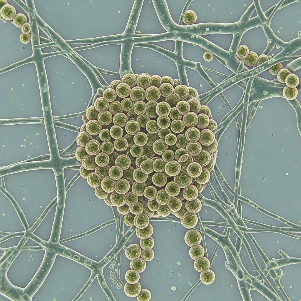
Aspergillosis encompasses a range of infections caused by Aspergillus species, ubiquitous molds found in soil, decaying vegetation, and indoor environments. While primarily affecting the lungs via inhalation of spores, cutaneous aspergillosis manifests as primary (direct skin inoculation) or secondary (disseminated from lungs or viscera) forms, predominantly in immunocompromised individuals.
What is aspergillosis?
Aspergillosis refers to diseases caused by fungi of the Aspergillus genus, with over 300 species identified, though A. fumigatus and A. flavus are most pathogenic in humans. These saprophytic molds thrive in organic matter and produce airborne conidia (spores) that enter the body primarily through inhalation. In healthy hosts, inhaled spores are cleared by alveolar macrophages; however, in immunocompromised patients—such as those with neutropenia, hematopoietic stem cell transplants, prolonged corticosteroid use, or AIDS—spores germinate into invasive hyphae, leading to angioinvasive disease. Cutaneous involvement occurs in 5-10% of disseminated cases, often signaling poor prognosis due to vascular invasion causing infarction. Primary cutaneous aspergillosis is rarer, resulting from direct inoculation at trauma sites, burns, surgical wounds, or intravenous catheter insertion points.
Who gets aspergillosis?
Immunocompetent individuals rarely develop invasive disease, experiencing at most allergic reactions like allergic bronchopulmonary aspergillosis (ABPA). High-risk groups for invasive and cutaneous aspergillosis include:
- Prolonged neutropenic patients (absolute neutrophil count <500/mm³ for >7 days)
- Allogeneic hematopoietic stem cell transplant recipients
- Solid organ transplant patients on immunosuppressants
- Patients with prolonged high-dose corticosteroids or TNF-α inhibitors
- Burn victims (>50% body surface area)
- Premature neonates in NICU with catheters or occlusive dressings
- Advanced AIDS (CD4 <50 cells/µL) or chronic granulomatous disease
Secondary cutaneous lesions arise from hematogenous dissemination, often from primary pulmonary infection, while primary forms link to local barrier breaches.
What causes aspergillosis?
Aspergillus fumigatus predominates in pulmonary and secondary cutaneous disease due to its thermotolerance and prolific sporulation. A. flavus is common in primary cutaneous cases, especially in burn wards and tropical regions. Pathogenesis involves spore inhalation or inoculation, germination (24-48 hours), hyphal invasion of endothelium, thrombosis, and tissue necrosis. Aspergillus’s angiotropism—invading vessels at 45° dichotomous branching—distinguishes it histologically. Virulence factors include gliotoxin (immunosuppressant), melanin (antioxidant), and proteases. Environmental exposure surges in construction, composting, or marijuana smoking.
What are the clinical features of aspergillosis?
Pulmonary aspergillosis
Manifests as invasive pulmonary aspergillosis (fever, dyspnea, hemoptysis, cavitary lesions on CT) or aspergilloma (fungus ball in pre-existing cavities).
Cutaneous aspergillosis
Primary cutaneous aspergillosis (1-3% of invasive cases) develops at inoculation sites:
- Initial erythema/induration evolving to necrotic eschar, hemorrhagic bullae, or ulcerative nodules
- Catheter sites: radial necrosis from puncture
- Burns: superficial spreading in macerated areas, ecthyma-like
- Neonates: granulation tissue or nodules
Secondary cutaneous aspergillosis (from dissemination):
- Multiple erythematous papules/macules progressing to necrotic papulonodules, bullae, or eschars
- Often trunk/extremities; mimics ecthyma gangrenosum or mucormycosis
- Paranasal sinus extension may cause facial cellulitis
| Type | Risk Factors | Skin Lesions | Prognosis |
|---|---|---|---|
| Primary | Burns, trauma, catheters, neonates | Necrotic eschar, bullae at site; single lesion | Good if excised early; dissemination risk high |
| Secondary | Neutropenia, transplants, dissemination from lungs | Multiple papulonodules, hemorrhagic bullae, eschars | Poor; >50% mortality |
Diagnosis of aspergillosis
Suspicion arises from risk factors + compatible lesions. Confirm via:
- Biopsy: Full-thickness to subcutis; shows 45° septate hyphae (GMS/PAS stain), angioinvasion, infarction, neutrophilic/granulomatous inflammation. Primary: superficial dermis; secondary: deep dermis/subcutis
- Culture: Sabouraud agar; identifies species (critical for A. terreus amphotericin resistance)
- Imaging: Chest CT halo/reverse halo signs for pulmonary source
- Biomarkers: Galactomannan (BAL/serum, sensitivity 80% in neutropenics), beta-D-glucan; PCR emerging
Differential: Mucormycosis (90° branching), Fusarium, Scedosporium, Pseudomonas ecthyma.
Treatment of aspergillosis
Urgent; reverse immunosuppression first. Tailor to type/site:
- Primary cutaneous: Surgical excision + antifungals (voriconazole 6 mg/kg IV BID day 1, then 4 mg/kg BID; or isavuconazole). Liposomal amphotericin B (5 mg/kg/day) alternative
- Secondary/invasive: Voriconazole first-line (preferred over amphotericin); duration 6-12 weeks minimum, guided by response
- Burns: Debridement/amputation + antifungals
- Prophylaxis: Posaconazole/itraconazole in high-risk hematology patients
Monitor LFTs (voriconazole hepatotoxicity); therapeutic drug monitoring essential.
What is the outcome for aspergillosis?
Primary cutaneous: >80% cure with early surgery/antifungals if non-disseminated. Invasive/secondary: 30-50% mortality, worse in prolonged neutropenia (>90% if >2 weeks). Burn aspergillosis: 80-100% mortality if >40% BSA involved. Recovery hinges on immune reconstitution.
Prevention of aspergillosis
- HEPA filtration, avoid construction dust in hospitals
- Antifungal prophylaxis (voriconazole/posaconazole) in high-risk
- Aseptic catheter care, minimize occlusive dressings
- Early neutropenia management
Frequently Asked Questions
What causes skin lesions in aspergillosis?
Skin lesions result from direct inoculation (primary) or hematogenous spread (secondary) of Aspergillus hyphae, which invade vessels causing necrosis.
Who is at highest risk for cutaneous aspergillosis?
Neutropenic patients, transplant recipients, burn victims (>50% BSA), and neonates with catheters.
How is cutaneous aspergillosis diagnosed?
By skin biopsy showing septate hyphae with 45° branching and culture confirmation.
What is the best treatment for aspergillosis skin infections?
Voriconazole is first-line; surgery for localized primary disease.
Can aspergillosis be prevented?
Yes, via antifungal prophylaxis in high-risk patients and environmental controls.
References
- Clinical Overview of Aspergillosis — Centers for Disease Control and Prevention (CDC). 2023-10-05. https://www.cdc.gov/aspergillosis/hcp/clinical-overview/index.html
- Diagnosis and Treatment of Cutaneous Aspergillosis — e-Journal of Medical Informatics. 2022-01-01. https://e-jmi.org/archive/detail/77?is_paper=y
- Cutaneous Aspergillosis — National Center for Biotechnology Information (NCBI) / PubMed Central. 2003-01-01. https://pmc.ncbi.nlm.nih.gov/articles/PMC105285/
- Aspergillosis — StatPearls / NCBI Bookshelf. 2023-07-17. https://www.ncbi.nlm.nih.gov/books/NBK482241/
- Fungus – Aspergillosis — Perri Dermatology. 2022-01-01. https://perridermatology.com/dr-perris-blog/fungus-aspergillosis/