
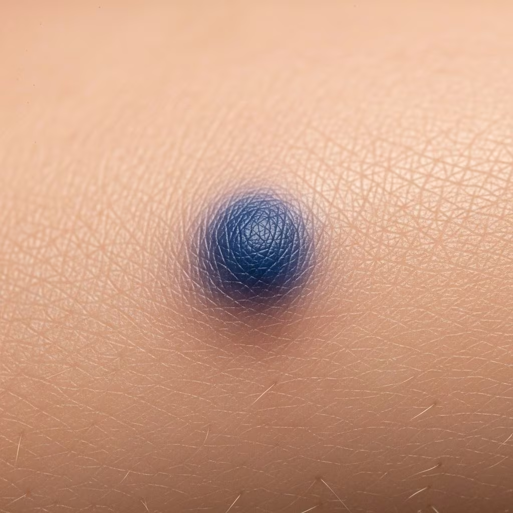
A blue naevus (nevus in American spelling) is a type of melanocytic naevus in which spindle-shaped or, less commonly, ovoid naevus cells are located most often wholly but at least partially within the dermis. These benign lesions derive their striking steel-blue colour from the deep location of melanin-producing cells in the skin, a phenomenon explained by the Tyndall effect where shorter blue wavelengths of light are scattered preferentially.
Who gets blue naevi?
Blue naevi are twice as common in women compared to men and more prevalent among Asians, with a rate of 3–5% versus 1–2% in other populations. They typically arise in late childhood or adolescence, though congenital blue naevi occur in about 1 in 3000 neonates, often becoming evident later. Common sites include the scalp, face, neck, dorsal hands and feet, buttocks, and sacral area. Eruptive forms may appear suddenly in immunosuppressed patients, during puberty, pregnancy, or after trauma like herpes zoster or sunburn.
What causes blue naevi?
Blue naevi result from the incomplete migration of melanocytes from the neural crest to the epidermis during fetal development. These dendritic melanocytes—bipolar or stellate cells with long processes—arrest in the dermis and produce abundant melanin. GNAQ/GNA11 mutations have been identified in various blue naevus subtypes, supporting their genetic basis. The deep dermal position causes the blue hue via the Tyndall effect, distinguishing them from superficial brown naevi.
What are the clinical features of blue naevus?
Blue naevi present as well-circumscribed, round or oval macules, papules, or nodules with uniform steel-blue, grey-blue, or bluish-black colour. Borders fade gradually into surrounding skin. Sizes vary: common types are 2–7 mm, slightly raised and smooth; cellular types reach 1–3 cm. They are usually solitary and stable lifelong, with no symptoms. Rare ectopic sites include oral mucosa, genitourinary tract, nails, lymph nodes, and prostate.
Main subtypes of blue naevus
- Common blue naevus: Small (2–7 mm), macular/papular/plaque-like, on head (esp. scalp), neck, hands/feet, sacral area. Benign, no malignant potential.
- Cellular blue naevus: Larger (1–3 cm), raised, on buttocks, sacral region, hands/feet. Higher cellularity; rare malignant transformation risk.
- Epithelioid blue naevus (pigmented epithelioid melanocytoma): Large epithelioid cells with pale cytoplasm, uniform nuclei, no mitoses.
- Plaque-type blue naevus: Extensive plaque form.
- Combined naevus: Mix of blue naevus with dysplastic, Spitz, or congenital elements.
Diagnosis of blue naevus
Diagnosis is primarily clinical based on typical appearance. Dermoscopy confirms with homogeneous steel-blue, hazy, or ground-glass pigmentation; rarely blue globules/dots. Scalp lesions or those >1 cm warrant caution due to monitoring difficulty. Histopathology shows spindle/epithelioid melanocytes in mid-to-deep dermis, sclerotic stroma, melanophages; positive SOX10/Melan-A stains. No epidermal involvement in pure forms.
Dermoscopy
The hallmark is uniform blue pigmentation. Exceptions include blue-brown globules/dots. This distinguishes from melanoma (asymmetric, multicoloured) or vascular lesions.
Differential diagnoses
Blue naevi mimic several lesions:
- Nodular melanoma: Irregular borders, colour variation, growth; biopsy essential.
- Dermatofibroma, venous lake, pilomatricoma, glomus tumour, angiokeratoma: Vascular or fibrous features on dermoscopy.
- Mongolian spots, naevus of Ota/Ito: Extensive dermal melanocytoses.
- Combined naevi: Mixed patterns.
Excisional biopsy resolves uncertainty, especially for de novo adult lesions, scalp sites, or changes.
Pathology of blue naevus
Histologically, blue naevi feature dermal dendritic melanocytes with abundant melanin, sclerotic stroma, melanophages, and periadnexal/neurovascular aggregation. Subtype specifics:
- Common: Slender dendrites, no epidermis involvement.
- Epithelioid: Large round cells, pale cytoplasm (post-bleach), uniform nuclei.
- Cellular: Dense oval/fusiform cells, deep lobular tongues into subcutis, multinucleate giants.
Mitoses suggest melanoma.
What is the treatment for blue naevus?
Most require no treatment as they are benign and stable. Excisional biopsy or removal for:
- Diagnostic uncertainty (e.g., >1 cm, adult onset, changing).
- Scalp location (hard to monitor).
- Cosmetic concerns.
Rare recurrence as satellites post-excision needs re-biopsy to exclude malignancy.
Outcome for blue naevus
Common blue naevi persist unchanged lifelong without complications. Cellular blue naevi rarely (<1%) transform to malignant melanoma; monitor for rapid growth/ulceration. Eruptive forms may regress spontaneously. Surgical removal prevents issues.
Frequently asked questions (FAQs) on blue naevus
Q: Is blue naevus cancerous?
A: Common types are benign. Cellular subtypes rarely become melanoma; monitor changes and biopsy if needed.
Q: Can blue naevus appear suddenly?
A: Yes, eruptive blue naevi occur rapidly in immunosuppressed patients, puberty, pregnancy, or post-trauma.
Q: Do blue naevi need removal?
A: Usually not, unless large, changing, on scalp, or cosmetic issue. Dermoscopy aids diagnosis.
Q: What does blue naevus look like under dermoscopy?
A: Homogeneous blue pigmentation, sometimes with globules/dots.
Q: Are blue naevi congenital?
A: Rare; most appear in childhood/adolescence. 1/3000 neonates affected.
Related topics
- Blue naevi pathology
- Blue naevus dermoscopy
- Melanocytic naevus
- Naevus of Ota
- Cellular blue naevus
References
- Blue naevi pathology — DermNet NZ. 2023. https://dermnetnz.org/topics/blue-naevi-pathology
- Blue naevus (blue nevus) — DermNet NZ. 2023. https://dermnetnz.org/topics/blue-naevus
- Blue naevus dermoscopy — DermNet NZ. 2023. https://dermnetnz.org/topics/blue-naevus-dermoscopy
- Blue Naevus (Causes, Symptoms, and Treatment) — Patient.info. 2024-01-15. https://patient.info/doctor/dermatology/blue-naevus
- Blue nevus on the scalp: Clinical and dermoscopic features — PMC (PubMed Central). 2024. https://pmc.ncbi.nlm.nih.gov/articles/PMC11141961/



