
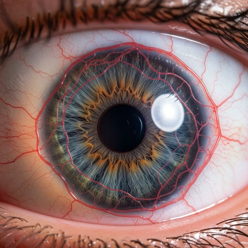
Corneal neovascularization (CNV) occurs when new, abnormal blood vessels grow into the normally avascular cornea, often triggered by injury, infection, or inflammation. This condition compromises corneal transparency and heightens risks for scarring, graft rejection, and vision loss. Early intervention is crucial to regress these vessels and restore ocular health.
Understanding the Pathophysiology of CNV
The cornea maintains clarity through its avascular structure, relying on tear film and aqueous humor for nutrients. CNV disrupts this balance when hypoxic or inflammatory signals activate pro-angiogenic factors like vascular endothelial growth factor (VEGF). These factors promote endothelial cell proliferation, migration, and tube formation, leading to leaky, immature vessels that cause edema and opacity.
Common triggers include trauma, chemical burns, infectious keratitis, contact lens overuse, and limbal stem cell deficiency. In post-keratoplasty eyes, CNV signals rejection risk. Imbalances between pro- and anti-angiogenic molecules drive progression, making targeted therapies essential.
Clinical Presentation and Diagnostic Approaches
Patients typically report blurred vision, photophobia, or foreign body sensation. Slit-lamp exam reveals superficial or deep stromal vessels, often with lipid deposition in chronic cases. Fluorescein angiography differentiates active from quiescent vessels, while anterior segment OCT quantifies vessel depth and density. Grading systems assess extent: peripheral (involving limbus), sectoral, or total corneal involvement.
- Superficial CNV: Linked to epithelial defects; responds well to topical agents.
- Deep stromal CNV: More resistant; may require invasive methods.
- Lymphatic vessels: Often coexist, contributing to immune-mediated rejection.
Pharmacological Interventions: First-Line Defenses
Anti-inflammatory and anti-angiogenic drugs form the cornerstone of CNV management, aiming to suppress vessel growth and promote regression.
Corticosteroids and NSAIDs
Topical corticosteroids like prednisolone acetate potently reduce inflammation and inhibit early angiogenesis. High-frequency dosing (e.g., hourly initially) controls acute CNV but shows limited efficacy on mature vessels. Nonsteroidal anti-inflammatory drugs (NSAIDs) like nepafenac provide adjunctive support by blocking prostaglandin-mediated vessel permeability.
Combination therapy maximizes outcomes: corticosteroids tackle inflammation while anti-VEGFs target angiogenesis. Risks include infection, glaucoma, and cataracts, necessitating close monitoring.
Anti-VEGF Therapies
Bevacizumab, ranibizumab, and aflibercept neutralize VEGF, regressing immature vessels. Topical formulations (1-2%) reduce vessel caliber and area, though penetration limits deep effects. Subconjunctival injections offer better bioavailability for superficial/mid-stromal CNV, while intrastromal delivery sustains levels in avascular stroma.
Studies show 40-70% vessel regression post-treatment, with ranibizumab narrowing stable vessels effectively. Deeper/mature CNV responds less, often requiring retreatment due to short half-lives.
Physical and Thermal Ablation Techniques
When drugs fail, direct vessel occlusion via heat or light provides rapid regression.
Laser Photocoagulation
Argon (514nm), yellow dye (577nm), and frequency-doubled Nd:YAG (532nm) lasers selectively coagulate hemoglobin-rich vessels. Outpatient procedures target efferent (wider, slower-flow) loops, sparing afferent feeders initially. One study reported 44% neovascular area reduction at 3 months, with 53% complete occlusion.
Combining laser with anti-VEGF mitigates VEGF upregulation from thermal injury. Complications include endothelial damage, hemorrhage, and recanalization (up to 37%).
Fine Needle Diathermy (FND)
This cost-effective method uses a 30-gauge needle to deliver low-heat current, cauterizing vessels at varying depths under topical anesthesia. It effectively closes both afferent and efferent segments, outperforming laser in some extensive cases. Retreatment rates are 20-30% due to reperfusion.
Adverse events: microperforation, hemorrhage, haze. FND suits resource-limited settings.
Advanced and Emerging Modalities
Photodynamic Therapy (PDT)
PDT employs verteporfin photosensitizer activated by laser (689nm), generating reactive oxygen species to occlude vessels selectively. Animal models confirm lymphatic regression with minimal adjacent damage, enhancing graft survival. Human trials show promise for refractory CNV.
Corneal Cross-Linking (CXL)
UVA-riboflavin CXL strengthens stroma and inhibits angiogenesis in burn models, reducing vessel extent versus controls. Extended irradiation yields superior results. Often combined with other therapies.
Immunomodulators and Novel Agents
Tocilizumab (IL-6 antagonist) matches bevacizumab efficacy subconjunctivally. Tyrosine kinase inhibitors and TNF-α blockers show preclinical success. Mitomycin C intravascular embolization halts vessel repair in lipid keratopathy.
Treatment Comparison: Pros, Cons, and Outcomes
| Treatment | Advantages | Limitations | Complications | Success Rate |
|---|---|---|---|---|
| Corticosteroids | Quick inflammation control; easy administration | Poor on mature vessels | Infection, glaucoma, cataract | 50-70% early regression |
| Anti-VEGF (topical/subconjunctival) | Targets active vessels; non-invasive options | Expensive; short duration | Epithelial defects, thinning | 40-70% area reduction |
| Laser Photocoagulation | Outpatient; precise | Recanalization; misses afferents | Endothelial damage, hemorrhage | 44-53% occlusion |
| Fine Needle Diathermy | Inexpensive; multi-depth | Needs retreatment | Perforation, haze | 60-80% initial closure |
| PDT/CXL | Selective; stromal strengthening | Experimental; access limited | Minimal reported | Promising in models |
Therapeutic Decision-Making: A Stepwise Approach
Tailor therapy to CNV extent, activity, and etiology. Start with topical steroids + anti-VEGF for active superficial CNV. Escalate to subconjunctival/intrastromal anti-VEGF or FND/laser for stromal involvement. Reserve PDT/CXL for refractory cases. Monitor quarterly with imaging; address underlying causes (e.g., dry eye, trichiasis).
- Acute/post-trauma: Aggressive steroids + topical anti-VEGF.
- Post-graft: Immunosuppressants + laser.
- Chronic/lipid-laden: FND + amniotic membrane.
Future Directions in CNV Management
Gene therapy silencing VEGF, nanoparticle drug delivery for sustained release, and stem cell therapies to rebuild limbal barriers hold potential. Clinical trials validate multi-target inhibitors. Personalized medicine via biomarker profiling (VEGF levels, vessel maturity) will optimize outcomes.
Frequently Asked Questions (FAQs)
What causes corneal neovascularization?
Inflammation, hypoxia, or injury disrupts angiogenic balance, spurring vessel ingrowth.
Can CNV be reversed completely?
Early intervention regresses 50-80% of vessels; mature ones may persist, requiring ongoing management.
Is anti-VEGF safe for long-term use?
Topical forms have favorable profiles; monitor for thinning. Subconjunctival safer than intravitreal.
How soon after diagnosis should treatment start?
Immediately for active CNV to prevent progression and scarring.
Does CNV affect contact lens wear?
Yes; halt use until regression to avoid exacerbation.
References
- Therapeutic approaches for corneal neovascularization — PMC/NCBI. 2017-11-15. https://pmc.ncbi.nlm.nih.gov/articles/PMC5723406/
- Management of corneal neovascularization: Current and emerging therapies — PMC/NCBI. 2015-11-20. https://pmc.ncbi.nlm.nih.gov/articles/PMC467007/
- Conquering Corneal Neovascularization — Corneal Physician. 2023-11-01. https://www.cornealphysician.com/issues/2023/november/conquering-corneal-neovascularization/
- Neovascularization of the Eye: Types & Treatment — Cleveland Clinic. 2023-05-10. https://my.clevelandclinic.org/health/diseases/24131-neovascularization-of-the-eye



