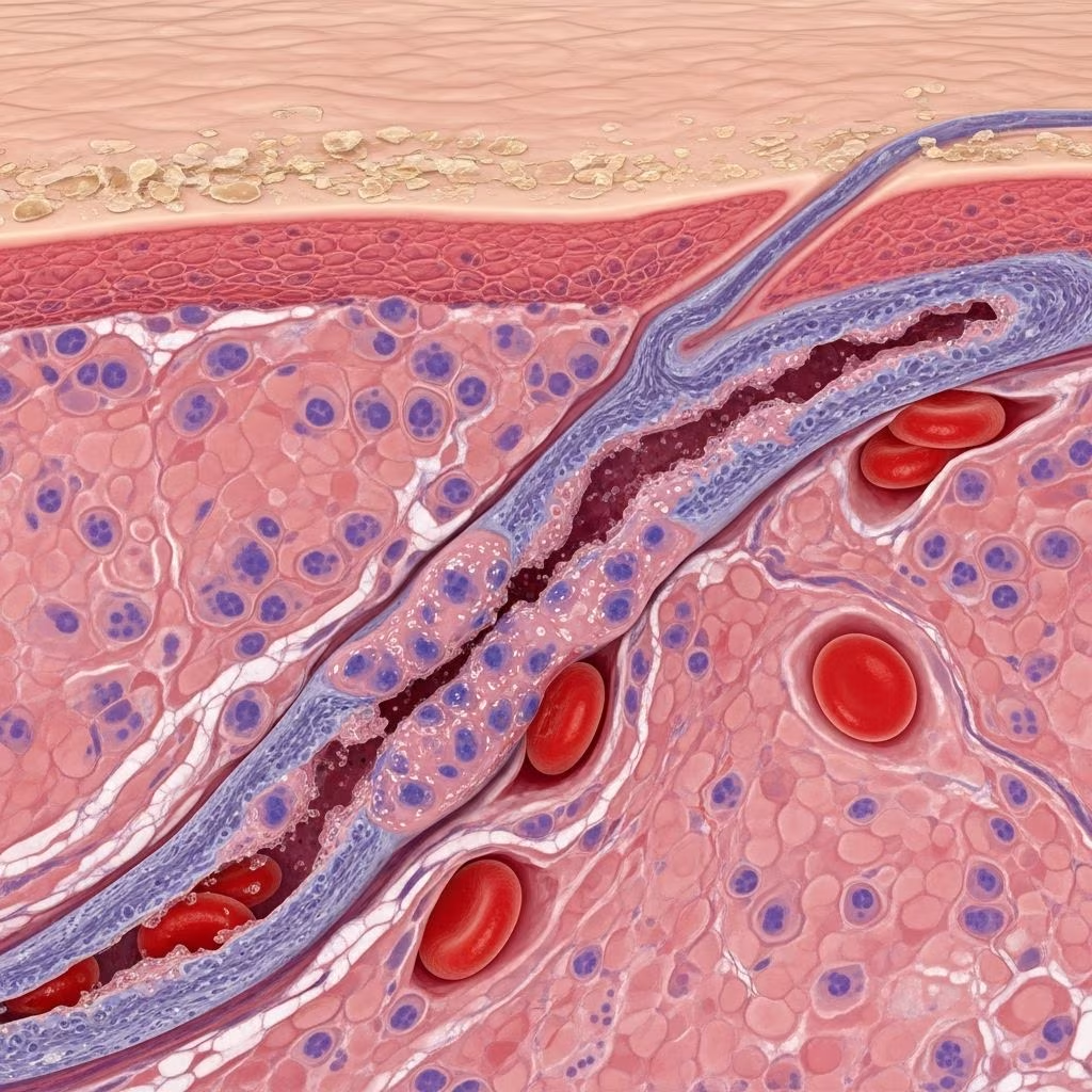
Cutaneous small vessel vasculitis(also known assmall vessel vasculitis,cutaneous leukocytoclastic angiitisorhypersensitivity vasculitis) is an inflammation of the small or post-capillary venules (<1 mm diameter) in the skin predominantly affecting thedermis.
What is small vessel vasculitis?
Vasculitisis a disorder in which there are inflamed blood vessels. These may include capillaries, arterioles, venules and lymphatics.
Small vessel vasculitisis the most common form of vasculitis affecting arterioles and venules.
Small vessel vasculitis is also called immune complex small vessel vasculitis.
The term hypersensitivity vasculitis is used for cutaneous small vessel vasculitis due to known drug or infection.
There are particular types of small vessel vasculitis that present with similar cutaneous signs and should be considered in the differential diagnosis. These include:
- IgA vasculitis(formerly Henoch 63Schlein purpura)
- Urticarial vasculitis
- Hypocomplementaemic vasculitis
- Cryoglobulinaemic vasculitis
Who gets cutaneous small vessel vasculitis (and why)?
Cutaneous small vessel vasculitis can affect any age but is uncommon in children and most often presents in the middle-aged and elderly.
In a large series of 440 patients over a 14-year period presenting with leukocytoclastic vasculitis of the skin, 82% had disease limited only to the skin, 12% had IgA vasculitis (Henoch 63Schlein purpura), and a small number of cases had an underlying systemic connective tissue disease.
Of those with cutaneous disease only, most cases were of unknown cause, 27% were drug-induced, 18% due to bacterial infection, 5% virally induced, and 1% had an underlying malignancy.
Causes of cutaneous small vessel vasculitis
The clinical features of hypersensitivity vasculitis include predominantlypalpable purpura(80 6390% of cases), usually after exposure to medications or infection.
Other skin lesions that may be present include:
- Urticaria(itchy weals)
- Erythema multiforme
- Bullae(blebs and blisters)
- Pustules
- Vesicles
- Ulcersandslowly healing erosions
Symptoms accompanying the skin lesions include:
- Pruritus(itching)
- Burning
- Pain
Drugs that may provoke small vessel vasculitis
Drug-induced small vessel vasculitis is the most common recognizable cause of cutaneous small vessel vasculitis.
| Drug class | Examples |
|---|---|
| Antibiotics | Sulfonamides, penicillins, cephalosporins, tetracyclines, minocycline, erythromycin, quinolones, isoniazid, rifampicin |
| NSAIDs | Ibuprofen, diclofenac, naproxen |
| Diuretics | Frusemide, hydrochlorothiazide, chlorthalidone |
| Antihypertensives | ACE inhibitors, calcium channel blockers |
| Others | Allopurinol, gold, granulocyte colony stimulating factor |
Vaccination-associated vasculitis has been rarely reported.
Infections
- Bacterial: Streptococcus, Staphylococcus, other respiratory tract infections
- Viral: hepatitis B and C, herpes simplex and zoster, influenza, HIV
- Mycoplasma, other respiratory tract pathogens
Systemic vasculitis
- ANCA-associated vasculitis
- Cryoglobulinaemia
Systemic diseases
- Connective tissue diseases: rheumatoid arthritis, Sjf6gren syndrome, systemic lupus erythematosus
- Sarcoidosis
Clinical features of cutaneous small vessel vasculitis
The initial acute rash of small vessel vasculitis usually subsides within 2 633 weeks, but crops of lesions may recur over weeks to several months, and hypersensitivity vasculitis may rarely become relapsing or chronic.
The most severe complications of small vessel vasculitis are seen with its systemic forms, such as kidney disease associated with IgA vasculitis.
Local ulceration from cutaneous small vessel vasculitis can lead to:
- Bacterial superinfection
- Milroy disease (extensive dry gangrene)

Purpuric rash due to cutaneous small vessel vasculitis
Diagnosis of cutaneous small vessel vasculitis
The clinical diagnosis of an acute cutaneous small vessel vasculitis is generally straightforward.
Thorough history and examination are essential to determine if symptoms and signs are confined to the skin, or if there may be systemic involvement, and to establish a cause.
Skin biopsy
Cutaneous small vessel vasculitis is confirmed by 4-mm punch biopsy of an early purpuric papule, ideally present for 24 6348 hours.
Histopathology reveals neutrophils around arterioles and venules, and fibrinoid necrosis (fibrin within or inside the vessel wall). There may be extravasated red cells, leukocytoclasis (broken-up neutrophils within the vessel wall) and signs of an underlying disease.
Blood tests
Initial investigations include:
- Full blood count
- Electrolytes, urea, creatinine
- Liver function
- Antinuclear antibody
- Rheumatoid factor
- C3, C4 complement
- Cryoglobulins
- Urine microscopy
- Antineutrophil cytoplasmic antibody
If an initial screen indicates an abnormality or there is clinical suspicion of a more widespread vasculitic process, further investigations will be requested.
The majority of patients presenting with palpable purpura have primary cutaneous small vessel vasculitis, and no underlying cause is found in spite of extensive investigations.
Management of cutaneous small vessel vasculitis
In most patients presenting with the first episode of acute cutaneous small vessel vasculitis, general measures are all that is required to keep the patient comfortable until the rash spontaneously resolves.
Symptomatic relief
- Rest and elevation of legs
- Cool compresses
- Paracetamol— for pain
- Topicalcorticosteroidsfor itch
Specific management
If a specific cause or association is identified, treat that (eg, discontinue offending medication or treat underlying infection).
Severe skin disease
- Prednisone0.5 631 mg/kg/day until rash settles
- Colchicine0.6 mg twice daily
- Dapsone50 63200 mg/day (avoid in G6PD deficiency)
Systemic vasculitis
Refer to rheumatologist, nephrologist, or other relevant specialist for management of systemic vasculitis.
What is the outlook for cutaneous small vessel vasculitis?
The acute rash of primary cutaneous small vessel vasculitis commonly resolves within 2 633 weeks, however it can take 6 6312 months to completely fade.
Recurrences are not uncommon but are usually milder than the initial flare.
Frequently asked questions
What does cutaneous small vessel vasculitis look like?
Cutaneous small vessel vasculitis mainly affects the lower legs and presents as palpable purpura (raised red or purple spots or patches) on both legs. Individual spots are usually 1 633 mm diameter but may coalesce to form larger areas of discolouration. The rash may be itchy or painful.
How is cutaneous small vessel vasculitis diagnosed?
The clinical diagnosis of an acute cutaneous small vessel vasculitis is generally straightforward. Thorough history and examination are essential. Cutaneous small vessel vasculitis is confirmed by 4-mm punch biopsy of an early purpuric papule, ideally present for 24 6348 hours.
How is cutaneous small vessel vasculitis treated?
In most patients presenting with the first episode of acute cutaneous small vessel vasculitis, general measures are all that is required to keep the patient comfortable until the rash spontaneously resolves. If a specific cause or association is identified, treat that (eg, discontinue offending medication or treat underlying infection).
References
- Cutaneous Small-Vessel Vasculitis 63 Vasculitis Foundation. 2024-02-05. https://vasculitisfoundation.org/education/vasculitis-types/cutaneous-small-vessel-vasculitis/
- Cutaneous small vessel vasculitis 63 PubMed. 2022-12-13. https://pubmed.ncbi.nlm.nih.gov/36524408/
- Cutaneous vasculitis; An algorithmic approach to diagnosis 63 Frontiers in Medicine. 2022-10-17. https://www.frontiersin.org/journals/medicine/articles/10.3389/fmed.2022.1012554/full
- Cutaneous Vasculitis Information for Patients 63 Rheumatology Dermatology Society. 2023. https://www.rheumaderm-society.org/vasculitis-information-for-patients/