
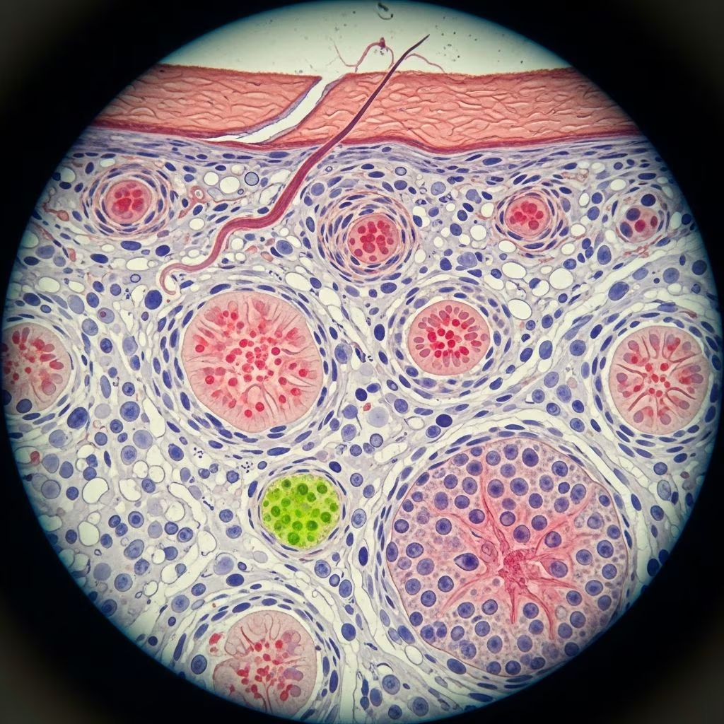
This comprehensive glossary outlines key terms used in dermatopathology, the microscopic study of skin diseases. Understanding these terms is crucial for interpreting skin biopsies accurately, distinguishing between inflammatory, neoplastic, and degenerative processes. Dermatopathologists rely on these descriptors to diagnose conditions ranging from eczema to melanoma.
Introduction
Dermatopathology involves examining skin tissue under a microscope to identify disease patterns. Terms describe changes in the epidermis, dermis, cellular alterations, and inflammatory patterns. These standardized descriptors ensure consistent communication among clinicians and pathologists worldwide. Common patterns include spongiotic dermatitis (eczema-like), psoriasiform (psoriasis-like), and lichenoid (interface) reactions.
Acantholysis
Acantholysis refers to the loss of cohesion between keratinocytes (epidermal cells), resulting in intraepidermal clefts or blisters. It occurs due to breakdown of desmosomal attachments. Classic examples include pemphigus vulgaris, where autoantibodies target desmogleins, leading to rounded acantholytic cells (“row of tombstones”). In Hailey-Hailey disease, genetic defects cause similar separation. Clinically, this manifests as flaccid blisters and erosions.
Acantholytic Dyskeratosis
Acantholytic dyskeratosis combines acantholysis with dyskeratosis (premature keratinization). Affected keratinocytes appear dysplastic with hyperchromatic nuclei and eosinophilic cytoplasm, forming ‘corps ronds’. Seen in Darier disease, warty dyskeratoma, and actinic keratosis. This focal change helps differentiate benign from premalignant lesions.
Acanthosis
Acanthosis is epidermal thickening due to increased spinous layer keratinocytes. It appears as elongation of rete ridges. Causes include chronic irritation (psoriasis, lichen simplex chronicus), obesity (acanthosis nigricans), or endocrine disorders. Regular acanthosis shows uniform rete elongation; irregular suggests neoplasia like squamous cell carcinoma.
Anagen
Anagen is the active growth phase of the hair cycle, lasting 2-6 years. Follicles produce hair shaft rapidly. Anagen effluvium, from chemotherapy, causes sudden hair loss by halting this phase. Histologically, anagen follicles are large with open hair shafts and prominent inner root sheath.
Apoptosis
Apoptosis (programmed cell death) appears as shrunken keratinocytes with pyknotic nuclei and eosinophilic cytoplasm, often in the basal layer. Seen in graft-versus-host disease, lichen planus, and viral infections. Clusters form ‘satellite cell necrosis’ when adjacent to damaged cells.
Atrophy
Atrophy denotes thinning of the epidermis or dermis. Epidermal atrophy shows flattened rete ridges; dermal from reduced collagen (steroid-induced, aging). In lichen sclerosus, atrophic epidermis overlies hyalinized dermis. Clinically, skin appears shiny and wrinkled.
Basement Membrane
The basement membrane is a thin acellular layer separating epidermis from dermis, visualized with PAS stain. Thickening occurs in dystrophic epidermolysis bullosa; duplication in lichen planus. It anchors epithelium and filters molecules.
Birefringence
Birefringence is the optical property where material splits polarized light, appearing bright against dark background. Useful for identifying amyloid (apple-green), collagen, or fibrin. Absent in necrosis, aiding differential diagnosis.
Bulla(e)
Bullae are large blisters (>0.5 cm) filled with fluid. Subepidermal (e.g., bullous pemphigoid) show intact epidermal roof; intraepidermal (pemphigus) have acantholytic bases. Tense bullae suggest subepidermal; flaccid intraepidermal.
Calcinosis Cutis
Calcinosis cutis involves dystrophic calcium deposition in damaged dermis. Basophilic granules or plates around vessels or eccrine ducts. Associated with scleroderma, lupus, or trauma. Dystrophic type follows inflammation; idiopathic is normocalcemic.
Catagen
Catagen is the involution phase of hair cycle (2-3 weeks), where follicle shrinks, apoptosis occurs, and bulb ascends. Seen in normal cycling or telogen effluvium triggers. Histology shows wrinkled outer root sheath.
Cornoid Lamella
Cornoid lamella is a thin parakeratotic column with absent granular layer beneath, overlying vacuolated keratinocytes. Hallmark of porokeratosis; also in actinic keratosis or squamous cell carcinoma.
Crust
Crust (scab) forms from dried serum, blood, or pus on skin surface. Histologically, eosinophilic with embedded neutrophils. Common in impetigo, infected eczema.
Cyst
Cyst is a cavity lined by epithelium containing keratin or fluid. Epidermoid cysts have stratified squamous lining; trichilemmal show abrupt keratinization. Rupture causes granulomatous inflammation.
Dermal Oedema
Dermal oedema widens collagen bundles with pale spaces. Acute in urticaria; chronic leads to fibrosis. Perivascular in insect bites.
Dermis
The dermis is connective tissue supporting epidermis, containing collagen, elastin, vessels, nerves. Superficial (papillary) is loose; reticular is dense. Solar damage affects both.
Dyskeratosis
Dyskeratosis is aberrant keratinization in lower epidermis, producing round eosinophilic cells (corps ronds, grains). Seen in viral warts, Bowen disease, Darier disease.
Elastosis
Elastosis (solar elastosis) is basophilic amorphous dermal material from sun-damaged elastic fibers. Amorphous on H&E; verified with elastic stain. Causes leathery skin in photoaging.
Flame Figures
Flame figures are eosinophilic deposits around hair follicles from degenerated eosinophils in eosinophilic cellulitis or Wells syndrome. Mimic Wells syndrome histologically.
Foam Cells
Foam cells are lipid-laden macrophages with foamy cytoplasm. Seen in xanthomas, dermatofibroma, or regressing melanomas.
Epidermis
The epidermis is stratified keratinized epithelium from basal to stratum corneum. Layers: basal, spinous, granular, corneum. Hyperplasia or atrophy alters thickness.
Epidermolytic Dyskeratosis
Epidermolytic dyskeratosis shows compact orthokeratosis, vacuolar keratinocytes, and clumped keratin filaments. Seen in epidermolytic ichthyosis, ichthyosis bullosa, palmoplantar keratoderma.
Epidermotropism
Epidermotropism is atypical lymphocytes migrating single-file into epidermis, hallmark of mycosis fungoides. Lacks spongiosis; forms Pautrier microabscesses.
Exocytosis
Exocytosis is inflammatory cells (lymphocytes, neutrophils) migrating into epidermis randomly. Common in spongiotic dermatitis like eczema.
Fibrinoid Necrosis
Fibrinoid necrosis is bright eosinophilic homogeneous material in vessel walls from fibrin deposition. Seen in vasculitis, collagen vascular diseases.
Fibrosis
Fibrosis is increased dermal collagen with reduced cellularity. Early sclerotic; late hyalinized. In scars, morphea, nephrogenic systemic fibrosis.
Follicular Plugging
Follicular plugging is keratin and debris filling follicles, forming comedones in acne or horny plugs in lupus erythematosus.
Granuloma
Granuloma is organized aggregate of epithelioid histiocytes, often with multinucleate giants. Foreign body type has polarizable material; palisaded in granuloma annulare.
Grenz Zone
Grenz zone is preserved compact dermis beneath subacute spongiotic epidermis, seen in early mycosis fungoides or insect bites.
Haemophagocytosis
Haemophagocytosis is macrophages engulfing blood cells, in histiocytic disorders or viral infections like EBV.
Haemosiderin Deposition
Haemosiderin deposition is golden-brown pigment from RBC breakdown, Prussian blue-positive. In stasis dermatitis, angiomas, amyloidosis.
Horn Cyst
Horn cyst is keratin-filled epidermal invagination, laminated. Seen in inverted follicular keratosis, trichilemmoma.
Hypergranulosis
Hypergranulosis is thickened granular layer, focal or diffuse, in warts, granular parakeratosis.
Hyperkeratosis
Hyperkeratosis is increased stratum corneum thickness. Orthokeratotic (no nuclei); parakeratotic (retained nuclei). In psoriasis, calluses.
Hyalinisation of Collagen
Hyalinisation of collagen is glassy homogenization of dermal collagen, pink on H&E. In lichen sclerosus, porphyria cutanea tarda.
Inflammation
Inflammation involves leukocyte infiltration responding to injury. Acute neutrophilic; chronic lymphocytic/histiocytic. Patterns: perivascular, lichenoid.
Interface Dermatitis
Interface dermatitis shows vacuolar basal layer change with apoptotic keratinocytes and lymphocytic infiltrate. In lupus, lichen planus, erythema multiforme.
Keratinisation
Keratinisation is terminal epidermal differentiation forming keratin. Aberrant in dyskeratosis.
Koilocytes
Koilocytes are HPV-infected keratinocytes with perinuclear halo, raisinoid nuclei. Hallmark of condyloma acuminatum.
Leucocytoclasia
Leucocytoclasia is karyorrhexis of neutrophils, in leukocytoclastic vasculitis with fibrinoid necrosis.
Lichenoid Reaction
Lichenoid reaction mimics lichen planus: band-like dermal lymphocytic infiltrate hugging epidermis, basal vacuolization. In drug eruptions, viral exanthems.
Mucinosis
Mucinosis is dermal mucopolysaccharide accumulation, Alcian blue-positive. In lupus, myxoid cysts, follicular mucinosis.
Necrobiosis
Necrobiosis is collagen degeneration with granulomatous response, eosinophilic swollen fibers. In granuloma annulare, necrobiosis lipoidica.
Necrosis
Necrosis is cell death with nuclear changes: pyknosis, karyorrhexis, karyolysis. Coagulative in infarcts; caseous in TB.
Neoplasm
Neoplasm is abnormal tissue growth, benign or malignant. Classified by origin: epithelial, melanocytic, mesenchymal.
Nodular Infiltrate
Nodular infiltrate is well-circumscribed dermal cellular aggregate, in lymphoma, metastases.
Oedema
Oedema is tissue fluid accumulation, pale spaces on histology. Clears with diastase.
Osteoma Cutis
Osteoma cutis is bone formation in skin, dystrophic post-trauma or progressive osseous heteroplasia.
Orthokeratosis
Orthokeratosis is basket-weave stratum corneum without nuclei, normal or thickened.
Pagetoid Spread
Pagetoid spread is intraepidermal malignant cell migration mimicking Paget disease, in melanoma, T-cell lymphoma.
Panniculus
Panniculus is subcutaneous fat layer, site of panniculitis.
Panniculitis
Panniculitis is septal or lobular fat inflammation, e.g., erythema nodosum (septal), pancreatic (lobular).
Papillary Mesenchymal Bodies
Papillary mesenchymal bodies are dilated capillaries with hyalinized stroma in papillary dermis, in regressing porokeratosis.
Papillomatosis
Papillomatosis is surface undulation with contiguous papillae, in warts, seborrheic keratosis.
Parakeratosis
Parakeratosis is stratum corneum with retained nuclei, in psoriasis, dermatophytosis.
Pautrier Microabscess
Pautrier microabscess is epidermal collection of atypical lymphocytes in mycosis fungoides.
Peri-adnexal Inflammation
Peri-adnexal inflammation surrounds follicles or glands, in rosacea, lupus.
Perivascular Lymphocytic Infiltrate
Perivascular lymphocytic infiltrate is superficial/deep lymphocytes around vessels, nonspecific in viral exanthems, early CTCL.
Pigment Incontinence
Pigment incontinence is melanin dropping into dermis from damaged basal cells, melanophages. In lichen planus, post-inflammation.
Pseudoepitheliomatous Hyperplasia
Pseudoepitheliomatous hyperplasia is reactive epidermal hyperplasia mimicking squamous cell carcinoma, in infections, halogenoderma.
Psoriasiform Hyperplasia
Psoriasiform hyperplasia shows regular acanthosis, thinned suprapapillary plates, parakeratosis. In psoriasis, Reiter syndrome.
Pustule
Pustule is intraepidermal neutrophil collection. Spongiform in pustular psoriasis; subcorneal in impetigo.
Pyknosis
Pyknosis is nuclear shrinkage in early necrosis/apoptosis.
Scale Crust
Scale crust combines parakeratosis, serum, inflammatory cells, in infected eczema.
Solar Elastosis
Solar elastosis is sun-induced dermal elastin/collagen degeneration, basophilic material.
Spongiosis
Spongiosis is intercellular edema causing epidermal spongelike appearance, in acute eczema.
Squamous Eddy
Squamous eddy is whorled keratinizing keratinocytes, in irritated seborrheic keratosis, squamous cell carcinoma.
Telogen
Telogen is resting hair phase (3 months), fibrous stremma, club hair. Effluvium shows increased telogen follicles.
Transepidermal Elimination
Transepidermal elimination extrudes dermal material (collagen, elastin) through epidermis, in perforating disorders like Kyrle disease.
Tumour
Tumour is solid neoplasm mass, from Latin ‘swelling’. Benign or malignant.
Vacuolar Degeneration
Vacuolar degeneration is basal keratinocyte cytoplasmic vacuoles progressing to apoptosis, in interface dermatitis.
Vasculitis
Vasculitis is vessel wall inflammation with fibrinoid necrosis, leukocytoclasia. Leukocytoclastic most common.
Vesicle
Vesicle is small blister (<0.5 cm). Similar to bulla but size-based.
Viral Inclusion Bodies
Viral inclusion bodies are intranuclear/cytoplasmic aggregates: eosinophilic (herpes), basophilic (HPV). Diagnostic clue.
Frequently Asked Questions (FAQs)
What is the difference between spongiosis and acantholysis?
Spongiosis is intercellular edema without cell separation; acantholysis involves loss of desmosomes causing clefts.
How does solar elastosis appear histologically?
As basophilic, amorphous dermal material with clumped elastic fibers on Verhoeff stain.
What causes Pautrier microabscesses?
Epidermotropism of atypical cerebriform lymphocytes in mycosis fungoides.
Is interface dermatitis always lichen planus?
No, also in lupus, drug eruptions, graft-versus-host disease.
What stain confirms haemosiderin?
Perls Prussian blue turns it blue.
References
- Dermatopathological terminology — DermNet New Zealand. 2023-10-15. https://dermnetnz.org/topics/dermatopathological-terminology
- Glossary of dermatopathological terms — BAD DermpathPRO Learning Hub. 2024-01-12. https://dermpathpro.com/glossary_of_terms/
- Dermatopathology: An Abridged Compendium — PMC (NCBI). 2013-05-29. https://pmc.ncbi.nlm.nih.gov/articles/PMC3663391/
- Dermatology Glossary of Terms — Robert Miller MD. 2022-08-05. http://www.robertmillermd.com/derma_glossary.html
- Dermatology Dictionary — PCDS. 2024-03-20. https://www.pcds.org.uk/dermatology-dictionary



