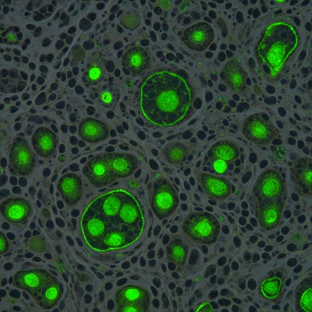
Direct immunofluorescence (DIF) is a specialized laboratory technique used primarily in dermatology to diagnose immune-mediated diseases affecting the skin, mucous membranes, kidneys, and other organs. It involves applying antibody-fluorophore conjugate molecules directly to fresh frozen tissue samples obtained from patient biopsies. These conjugates bind to specific abnormal protein depositions, such as immunoglobulins or complement components, in the tissue. Under a fluorescence microscope, the fluorophore emits light at a specific wavelength, revealing the location, pattern, and type of immune deposits. This visual information, combined with clinical history and routine histology, enables precise diagnosis of conditions like autoimmune bullous diseases, connective tissue disorders, and vasculitis.
What is direct immunofluorescence?
DIF detects in vivo deposited immunoreactants—including IgG, IgA, IgM, C3, fibrinogen, and other proteins—in patient tissue. Unlike indirect immunofluorescence (IIF), which uses patient serum on a substrate to detect circulating antibodies, DIF is a single-step process applied directly to the biopsy specimen. The technique is particularly valuable because it localizes immune complexes at sites like the dermo-epidermal junction, intercellular spaces, or blood vessel walls, providing diagnostic specificity. Studies show high concordance rates, with DIF positivity confirming diagnoses in up to 98% of immune-mediated skin disorders when correlated with histopathology.
The process requires fresh tissue snap-frozen in liquid nitrogen or Michel’s transport medium to preserve antigens. Paraffin-embedded tissue can be used after antigen retrieval (paraffin IF), but sensitivity drops to 0-79% compared to fresh samples. Pathologists analyze slides for deposition sites, immunoglobulin classes, and patterns, classifying them into linear, granular, or other morphologies.
Indications for direct immunofluorescence
DIF is indicated when immune-mediated diseases are suspected based on clinical and histopathological findings. It confirms diagnoses in bullous diseases, vascular inflammatory conditions, and connective tissue disorders, often providing definitive evidence where routine histology is inconclusive. DIF should always be interpreted alongside H&E-stained sections and patient history for optimal diagnostic yield. It is not useful for eczematous dermatitis or non-immune conditions.
Autoimmune bullous diseases
DIF is cornerstone for diagnosing autoimmune bullous diseases (AIBDs), where tissue-bound autoantibodies cause blistering. Common indications include:
- Pemphigus vulgaris and variants (foliaceus, paraneoplastic): Intercellular IgG and C3 deposition in the epidermis (fishnet pattern).
- Bullous pemphigoid (all forms, including gestationis): Linear IgG and C3 at the dermo-epidermal junction. DIF positive in 96-98% of cases.
- Dermatitis herpetiformis: Granular IgA in dermal papillae.
- Linear IgA bullous disease: Linear IgA at basement membrane.
- Epidermolysis bullosa acquisita: Linear IgG/C3 at DEJ, distinguished by salt-split skin in IIF.
- Intercellular IgA dermatosis: IgA in epidermal intercellular spaces.
In a study of 215 biopsies, DIF achieved 98.1% sensitivity in pemphigus and 96% in bullous pemphigoid, with negative results ruling out AIBDs effectively.
Connective tissue diseases
DIF aids in diagnosing:
- Lupus erythematosus (LE): Lupus band test—granular IgG, IgM, C3 at DEJ in lesional (100% positive) and non-lesional skin. DIF positive in 100% of LE cases.
- Dermatomyositis: Continuous C3 and IgG at DEJ.
Vasculitis and other inflammatory diseases
DIF identifies immune complex vasculitis and related conditions:
- Henoch-Schönlein purpura (IgA vasculitis): IgA in dermal vessels (100% DIF positive).
- Leukocytoclastic vasculitis: C3, fibrinogen in vessels.
- Essential mixed cryoglobulinemia: IgM/IgG in vessels.
- Polyarteritis nodosa: Fibrinogen, C3 in vessel walls.
Table 1 summarizes common indications:
| Category | Diseases |
|---|---|
| Bullous diseases | Pemphigoid (all forms), Pemphigus (all forms), Dermatitis herpetiformis, Linear IgA disease, Epidermolysis bullosa acquisita, Intercellular IgA dermatosis |
| Vascular diseases | Henoch-Schönlein purpura, Leukocytoclastic vasculitis, Essential mixed cryoglobulinemia, Polyarteritis nodosa |
| Connective tissue | Lupus erythematosus, Dermatomyositis |
Biopsy site and technique
Optimal biopsy site varies by disease to maximize diagnostic yield. Perilesional skin (2-3 mm from lesion edge) is preferred for most bullous diseases to avoid false negatives from inflammation.
| Disease | Optimal Biopsy Site |
|---|---|
| Pemphigus | Lesion edge or floor |
| Bullous pemphigoid | Small vesicle edge |
| Dermatitis herpetiformis | Intact blister or erythematous border |
| Lupus erythematosus | Non-sun-exposed normal skin for lupus band test |
| Vasculitis | Erythematous/active border of new lesion (<24h old); avoid ulcers/old lesions |
| Porphyria cutanea tarda | Involved skin (avoid ulcers/old lesions) |
A separate biopsy for H&E histology is routine, as DIF and histology require different processing. DIF uses frozen sections; process promptly in transport medium. DIF may be manual or automated.
Method of direct immunofluorescence
- Tissue preparation: Snap-freeze biopsy in liquid nitrogen or fix in Michel’s medium; cut 4-6 μm frozen sections.
- Staining: Incubate sections with fluorescein-conjugated antisera (anti-IgG, IgA, IgM, C3, fibrinogen).
- Washing and mounting: Rinse excess antibody; mount in anti-fade medium.
- Examination: View under fluorescence microscope (FITC filter); note intensity (0-4+), location, pattern.
Multiple fluorophores allow simultaneous detection (e.g., FITC for IgG, rhodamine for C3).
Interpretation of results
Pathologists classify findings by:
- Primary site: DEJ, intercellular, dermal vessels, perivascular, periadnexal.
- Immunoreactants: IgG (most common), IgA, IgM, C3, fibrinogen.
- Patterns:
- Linear (e.g., bullous pemphigoid).
- Granular (e.g., dermatitis herpetiformis, LE).
- Fishnet/intercellular (pemphigus).
- Vascular (vasculitis).
Five main groups: epidermal intercellular, basement membrane zone linear/granular, dermal papillary, vascular, deep dermal. Intensity correlates with disease activity in some AIBDs. Negative DIF excludes immune-mediated disease in 93.4% concordance with clinical/histology.
Frequently Asked Questions (FAQs)
What is the sensitivity of DIF in bullous pemphigoid?
DIF shows 96% positivity in bullous pemphigoid cases, with linear IgG/C3 at DEJ.
Is DIF useful for dermatitis herpetiformis?
Yes, granular IgA in dermal papillae is diagnostic.
Can DIF be done on paraffin tissue?
Yes (paraffin IF), but less sensitive (0-79%) than frozen sections.
What biopsy site for vasculitis DIF?
Erythematous border of lesion <24 hours old.
Does DIF replace histology?
No; best used with H&E and clinical correlation.
References
- Direct immunofluorescence – DermNet — DermNet NZ. 2023. https://dermnetnz.org/topics/direct-immunofluorescence
- Direct Immunofluorescence – PMC – NIH — National Library of Medicine. 2024-03-15. https://pmc.ncbi.nlm.nih.gov/articles/PMC10919951/
- Role of direct immunofluorescence in dermatological disorders — PubMed. 2015-06. https://pubmed.ncbi.nlm.nih.gov/26009711/
- Immunofluorescence in dermatology: A brief review — Journal of Skin and Sexually Transmitted Diseases. 2023. https://jsstd.org/immunofluorescence-in-dermatology-a-brief-review/
- Immunofluorescence in dermatology — Indian Journal of Dermatology, Venereology and Leprology. 2018. https://ijdvl.com/immunofluorescence-in-dermatology/
- Direct and Indirect Immunofluorescent Services — Weill Cornell Medicine Dermatopathology. 2024. https://dermpath.weill.cornell.edu/clinicians/direct-and-indirect-immunofluorescent-services