
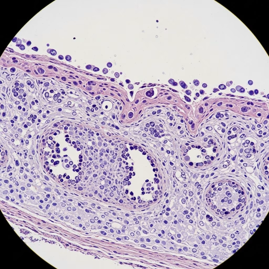
Erythema multiforme (EM) represents a prototypical type IV hypersensitivity reaction characterised by acute, self-limiting mucocutaneous lesions, most frequently triggered by herpes simplex virus (HSV) infection. This article dissects the intricate histological patterns and immunological mechanisms driving EM, emphasising the pivotal role of cytotoxic T cells targeting keratinocytes harbouring viral antigens.
What is Erythema Multiforme?
Erythema multiforme manifests as polymorphic skin eruptions featuring targetoid lesions with concentric zones of erythema, pallor, and central duskiness or blistering, predominantly on acral sites. It encompasses EM minor (skin-limited) and EM major (with mucosal involvement), distinguishing it from severe entities like Stevens-Johnson syndrome/toxic epidermal necrolysis (SJS/TEN) spectrum disorders.
- Classic morphology: Attollens (raised) target lesions with three zones – outer erythematous halo, pale edematous ring, central purpura/vesicle.
- Distribution: Symmetrical acral predominance (dorsal hands, forearms, feet, elbows, knees).
- Mucosal involvement: Oral erosions in 25-60% of recurrent HSV-associated cases.
EM typically affects young adults (20-40 years), with males twice as likely as females. Prodromal symptoms are absent in minor forms but may precede major variants.
Aetiology and Triggers
Over 90% of EM cases link to infections, with HSV-1 implicated in 70-80% of recurrent episodes. Other triggers include Mycoplasma pneumoniae (5-15%), medications (10-20%), and rarely autoimmune diseases or malignancies.
| Trigger | Frequency | Key Associations |
|---|---|---|
| HSV-1/2 | 70-80% | Recurrent EM, acral targets |
| Mycoplasma pneumoniae | 5-15% | Mucosal EM major, prodrome |
| Drugs (e.g., sulfonamides, anticonvulsants) | 10-20% | Single episode, atypical targets |
| Other infections (fungal, EBV) | <5% | Idiopathic/recurrent |
Genetic predisposition via HLA alleles (DQ3, DR53, B35) heightens susceptibility, particularly for post-HSV recurrences.
Pathogenesis: Immune Mechanisms
EM exemplifies cell-mediated cytotoxicity wherein CD8+ T lymphocytes recognise HSV-derived antigens on keratinocytes, unleashing a cytokine storm culminating in apoptotic epidermal necrosis. Absent infectious viral replication, EM arises from hypersensitivity to persistent viral DNA fragments.
HSV-Associated Mechanism
Post-HSV replication, basal keratinocytes synthesise viral DNA polymerase (Pol), presenting neoantigens via MHC class I to circulating CD8+ T cells. These effector cells, primed by prior infection, infiltrate lesional skin, releasing perforin/granzyme B and cytokines (IFN-γ, TNF-α), inducing keratinocyte apoptosis.
- Key steps:
- Monocytes transport HSV DNA fragments to distant keratinocytes.
- CD4+ Th1 cells amplify response via IFN-γ.
- CD8+ cytotoxic T cells target MHC I-presented viral peptides.
- Cytokine milieu (IFN-γ dominant) drives basal vacuolisation and full-thickness necrosis.
100% of HSV-EM biopsies detect viral Pol DNA exclusively in keratinocytes, confirming post-viral hypersensitivity sans active viraemia.
Drug-Induced Variant
Divergent from HSV-EM, drug reactions feature TNF-α predominance over IFN-γ, with earlier keratinocyte necrosis mediated by CD8+ cells recognising haptenised proteins. Necrotic keratinocytes colocalise with TNF-α secreting cells.
Mycoplasma-Triggered EM
Molecular mimicry between Mycoplasma antigens and keratinocyte self-proteins provokes autoreactive T cells, mirroring HSV pathways but with pulmonary prodrome.
Histological Features
Skin biopsy remains gold standard, revealing interface vacuolar dermatitis with lymphocyte satellitosis around apoptotic keratinocytes – the hallmark “satellite cell necrosis”.
Early Lesion (6-24 hours)
- Dermoepidermal junction: Vacuolar degeneration of basal layer, colloid bodies (Civatte bodies).
- Lymphocytic infiltrate: Predominantly CD8+ T cells and macrophages hugging basal keratinocytes (satellitosis).
- Cytokines: IFN-γ expression maximal.
Fully Developed Lesion (24-72 hours)
- Epidermis: Confluent keratinocyte necrosis, subepidermal clefting, intraepidermal vesicles.
- Dermis: Superficial perivascular lymphocytic infiltrate (CD4+/CD8+), papillary dermal edema.
- Apoptosis: Full-thickness dyskeratosis with hypereosinophilic keratinocytes.
Direct immunofluorescence (DIF) discloses granular C3/C1q deposition at DEJ in 50% HSV-EM cases, absent in drug reactions.
| Feature | HSV-EM | Drug-EM | SJS/TEN |
|---|---|---|---|
| Satellitosis | Prominent | Mild | Absent |
| Dominant cytokine | IFN-γ | TNF-α | FasL/Granzyme |
| DEJ vacuolisation | Focal | Diffuse | Full-thickness |
| Viral DNA PCR | Positive (keratinocytes) | Negative | Negative |
Mucosal Biopsies
Mirror cutaneous findings but with intensified epithelial sloughing and neutrophilic crypt abscesses in severe EM major.
Diagnostic Confirmation
Integrate clinical morphology, history, and biopsy:
- PCR: HSV DNA in lesional keratinocytes (96% sensitivity).
- IHC: CD8+ satellitosis, MxA (IFN-α induced).
- Serology: Mycoplasma IgM if pulmonary symptoms.
Differentiate from urticaria multiforme (young children, resolves 24h), rowell syndrome (lupus+EM), or erythema gyratum.
Frequently Asked Questions (FAQs)
Q: What causes the target lesions in erythema multiforme?
A: Cytotoxic CD8+ T cells induce keratinocyte apoptosis via perforin/granzyme, creating concentric zones of edema and necrosis.
Q: How does HSV trigger EM without active infection?
A: Keratinocytes express HSV DNA polymerase, presented via MHC I to memory T cells, provoking hypersensitivity.
Q: Can biopsy distinguish HSV-EM from drug-induced EM?
A: Yes – IFN-γ/CD8+ satellitosis favours HSV; TNF-α/earlier necrosis suggests drugs. PCR confirms HSV DNA.
Q: Is erythema multiforme contagious?
A: No, it is a sterile hypersensitivity reaction; contagion risk stems solely from underlying HSV.
Q: What HLA types predispose to recurrent EM?
A: DQ3, DR4, DR53, B35 strongly associated with post-HSV recurrences.
Clinical Implications and Management Insights
Understanding these mechanisms guides prophylaxis: valacyclovir prevents 50% HSV-EM recurrences. Avoid implicated drugs in drug-EM. Biopsy early lesions maximises diagnostic yield.
Recent advances highlight IL-15 upregulation in keratinocytes amplifying T-cell cytotoxicity, potential therapeutic target.
References
- Erythema multiforme — Wikipedia. 2024-01-15. https://en.wikipedia.org/wiki/Erythema_multiforme
- Erythema Multiforme — StatPearls, NCBI Bookshelf. 2023-10-01. https://www.ncbi.nlm.nih.gov/books/NBK470259/
- Erythema Multiforme | Symptoms, Signs — Geeky Medics. 2024-05-20. https://geekymedics.com/erythema-multiforme/
- Correlations Between Clinical Patterns and Causes of Erythema… — JAMA Dermatology. 2005-06-01. https://jamanetwork.com/journals/jamadermatology/fullarticle/478935
- Pathogenesis, clinical features, and diagnosis of erythema multiforme — UpToDate. 2024-11-10. https://doctorabad.com/uptodate/d/topic.htm?path=pathogenesis-clinical-features-and-diagnosis-of-erythema-multiforme



