
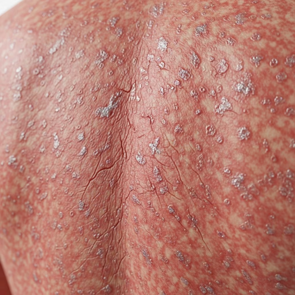
Erythroderma (also known as exfoliative dermatitis) is a generalised inflammatory skin condition characterised by erythema (redness) involving more than 90% of the body surface, with variable scaling, oedema and pruritus. It is a clinical syndrome rather than a specific disease and may arise from a wide range of underlying dermatoses, drugs or malignancies. Erythroderma represents a dermatological emergency due to risks of fluid loss, thermoregulatory disturbance, high-output cardiac failure, infection and death. Prompt diagnosis and management of the underlying cause is essential.
What is the cause of erythroderma?
Erythroderma may be primary (due to a dermatosis) or secondary (to drugs, malignancy or other systemic disorder). The most common causes vary by geographical region and patient population.
Primary causes
- Psoriasis: Accounts for 25–75% of cases in Western countries. Erythrodermic psoriasis often arises in patients with chronic plaque psoriasis following abrupt withdrawal of systemic therapy (e.g. potent topical steroids, methotrexate), infections or alcohol excess.
- Atopic dermatitis: Common cause in children and young adults, comprising 10–20% of cases. Widespread erythema with accentuated flexural involvement and fine scaling.
- Contact dermatitis: Allergic or irritant, often affecting 5–10% of cases.
- Pityriasis rubra pilaris: Features orange palmoplantar keratoderma and islands of sparing (1–8%).
- Seborrhoeic dermatitis: Rare cause.
- Lichen planus, bullous disorders (pemphigus foliaceus, pemphigus vulgaris), paraneoplastic pemphigus.
Secondary causes
- Drugs: Most common secondary cause (15–30%). High-risk drugs include anticonvulsants (carbamazepine, phenytoin, phenobarbitone), allopurinol, sulfonamides, β-lactam antibiotics, proton pump inhibitors and antitubercular drugs. Onset typically 1–6 weeks after drug initiation.
- Cutaneous T-cell lymphoma (CTCL): Including Sézary syndrome (5–10%).
- Other malignancies: Solid tumours, leukaemia (rare).
Idiopathic cases (10–30%) have no identifiable cause after thorough investigation.
Who gets erythroderma?
Erythroderma affects all ages but is more common in males (M:F = 3:1) and adults over 50 years. Incidence is 1–2 per 10,000 but may be underreported. Pre-existing inflammatory dermatoses increase risk.
What are the clinical features of erythroderma?
Acute phase (days–weeks): Rapid onset of generalised bright red erythema with early fine scaling. Oedema of face, hands and feet. Pruritus (intense), pain and chills.
Subacute/chronic phase: Thickened adherent scaling, fissuring, nail dystrophy (onycholysis, subungual hyperkeratosis), alopecia and ectropion.
Skin features
- Generalised erythema >90% body surface area
- Scaling: fine (psoriasis, atopy), coarse (CTCL), exfoliative
- Islands of normal skin: psoriasis (25%), pityriasis rubra pilaris
- Palmoplantar keratoderma: pityriasis rubra pilaris
Systemic features
| System | Manifestations |
|---|---|
| Cardiovascular | High-output cardiac failure (10–20%), oedema |
| Thermoregulatory | Hypothermia (<35°C), hyperthermia |
| Fluid/electrolyte | Dehydration, hypoproteinaemia (<50 g/L), hyponatraemia |
| Haematological | Anaemia, leucocytosis/eosinophilia, thrombocytosis |
| Metabolic | Negative nitrogen balance, hyperuricaemia |
How is erythroderma diagnosed?
Diagnosis is clinical but requires exclusion of underlying cause via history, examination, blood tests, skin biopsy(ies) ± other investigations.
History
- Preceding dermatosis, recent drugs (past 2 months), infections, malignancy
- Systemic symptoms, alcohol excess, medication non-compliance
Examination
- Mucosal involvement (CTCL, drugs), lymphadenopathy (CTCL), hepatosplenomegaly
- Palmoplantar involvement, nail changes, scalp involvement
Investigations
- Blood tests: FBC, U&E, LFT, albumin, CRP, uric acid, Sézary count (if CTCL suspected)
- Skin biopsy: Often multiple (1–3 sites). Features nonspecific but help narrow differentials:
- Psoriasis: regular acanthosis, parakeratosis, Munro microabscesses
- Drug: interface dermatitis, eosinophils
- CTCL: epidermotropism, atypical lymphocytes
- Other: Skin swab (infection), T-cell receptor gene rearrangement (persistent cases), lymph node biopsy
What is the differential diagnosis for erythroderma?
- Generalised exfoliative drug eruption
- Staphylococcal scalded skin syndrome (children)
- Toxic epidermal necrolysis (mucosal involvement)
- Generalised pustular psoriasis
- Sweet syndrome (neutrophilic)
Complications of erythroderma
- Infection: Bacterial (Staphylococcus aureus sepsis), viral, fungal (5–30%)
- High-output cardiac failure (10%)
- Thermoregulatory failure
- Thromboembolism, renal failure
- Mortality: 20–40% in elderly; lower (<10%) if cause addressed promptly
How is erythroderma treated?
Management is supportive + cause-directed. Admission to HDU/ICU often required.
General measures
- Fluids/electrolytes: Monitor insensible losses (4–5 L/day), IV fluids guided by U&E
- Nutrition: High protein (1.5–2 g/kg/day), enteral preferred
- Temperature: Warm environment (28–32°C), space blanket
- Skin care: Emollients (50:50 paraffin), wet wraps, avoid irritants
- Infection prophylaxis: Topical/systemic antibiotics if indicated
- Eye care: Lubricants for ectropion
Cause-specific therapy
| Cause | Treatment |
|---|---|
| Psoriasis | Cyclosporin (3–5 mg/kg/day) or infliximab (5 mg/kg) first-line for unstable cases; acitretin/methotrexate for stable |
| Atopic dermatitis | TCI/medium potency TCS + wet wraps, ciclosporin |
| Drugs | Drug cessation + supportive; steroids cautious |
| CTCL |
Avoid systemic steroids unless life-threatening (risk of rebound psoriasis).
Prevention of erythroderma
- Avoid triggers in high-risk patients (abrupt therapy withdrawal)
- Drug allergy documentation/bracelet
- Patient education on early warning signs
Prognosis of erythroderma
Mortality 10–40% (higher in elderly/malignancy). Good prognosis if cause reversible (psoriasis, drugs). Recurrence common (20–50%) if underlying disease uncontrolled.
Frequently asked questions
Q: Is erythroderma contagious?
A: No. It is not infectious.
Q: How long does erythroderma last?
A: 2–6 weeks with treatment; longer if idiopathic/malignant.
Q: Can erythroderma be fatal?
A: Yes, due to complications (infection, cardiac failure).
References and Further Reading
Books
- Rook’s Textbook of Dermatology
- Fitzpatrick’s Dermatology
Patient information
References
- Erythrodermic psoriasis: pathophysiology and current treatment strategies — National Psoriasis Foundation. 2017-08-01. https://pmc.ncbi.nlm.nih.gov/articles/PMC5572467/
- A Practical Approach to the Diagnosis and Treatment of Adult Erythroderma — Actas Dermo-Sifiliográficas. 2019-01-15. https://www.actasdermo.org/es-a-practical-approach-diagnosis-treatment-articulo-S1578219018303536
- Erythroderma — DermNet NZ. 2023-05-12. https://dermnetnz.org/topics/erythroderma



