
What is erythrokeratoderma?
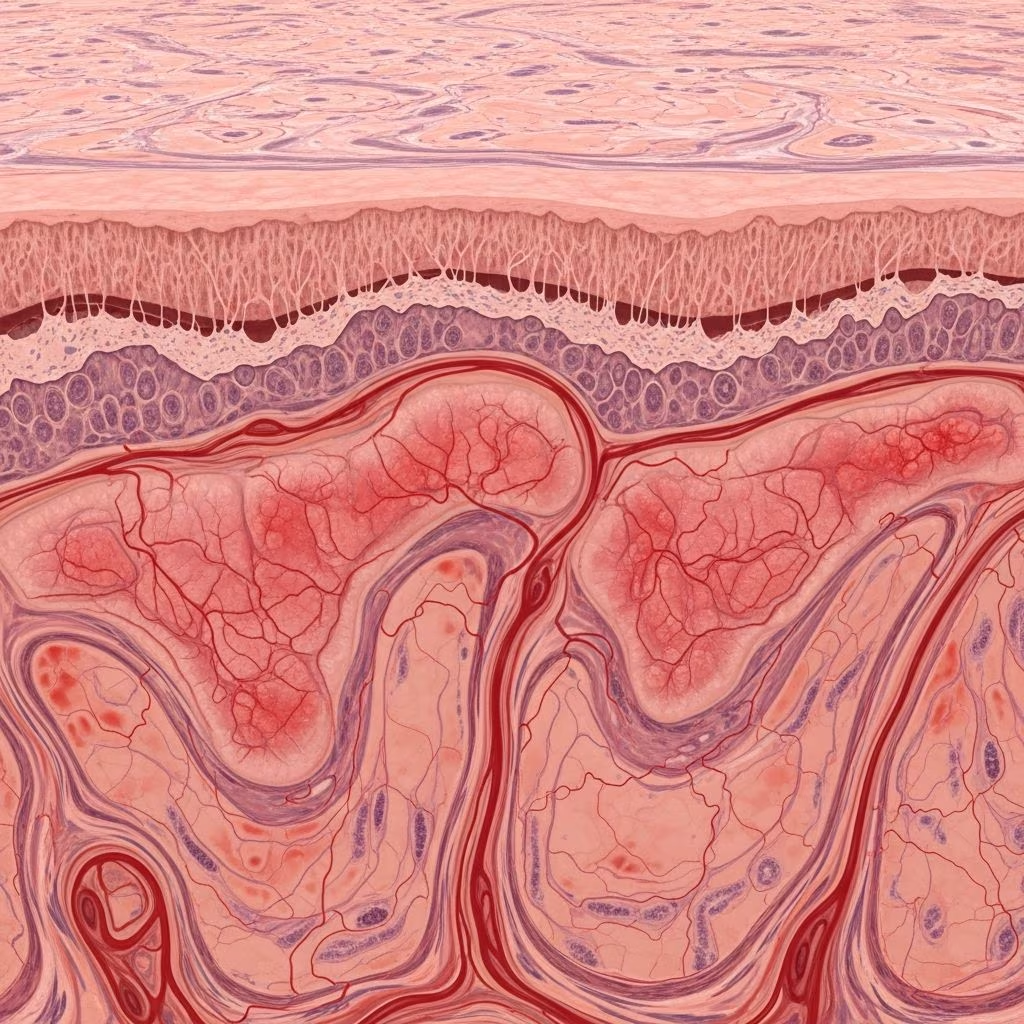
Erythrokeratoderma, sometimes referred to as erythrokeratodermia, describes a rare group of genetic disorders of keratinisation—the process forming the epidermis layers. These conditions feature well-demarcated plaques of erythema (redness) and hyperkeratosis (scaling). Affected skin shows persistent or transient red patches alongside thickened, scaly areas, varying by subtype.
Prevalence is extremely low, often familial, with onset at birth or early infancy. Physical and mental development remains normal, distinguishing it from broader syndromes. Severity fluctuates, even within families, impacting quality of life through cosmetic concerns, itching, or discomfort.
Who gets erythrokeratoderma?
These disorders are inherited, primarily autosomal dominant, affecting males and females equally. Erythrokeratoderma variabilis (EKV) often appears in infancy, while progressive symmetric erythrokeratoderma (PSEK) starts shortly after birth. Rare mosaic forms limit lesions to one body side.
- EKV: Present at birth or within first year; common in families with GJB3 or GJB4 mutations.
- PSEK: Infants develop symmetric plaques on face, limbs, buttocks.
- Other variants: Associated with KDSR mutations or cardiomyopathy in specific cases.
Geographic distribution shows no strong ethnic bias, though reported cases cluster in Europe and North America due to diagnostic access.
What causes erythrokeratoderma?
Genetic mutations disrupt skin barrier proteins, leading to abnormal cornification. Key genes include:
| Subtype | Gene | Protein | Inheritance |
|---|---|---|---|
| EKV | GJB3 (connexin 31), GJB4 (connexin 30.3) | Gap junctions | Autosomal dominant |
| PSEK | KDSR | Ceramide synthase | Autosomal dominant/somatic |
| EKVP | GJB3/GJB4 | Gap junctions | Autosomal dominant |
| Cardiomyopathy-associated | DSC1/DSC2 | Desmoglein | Sporadic |
Mutations impair intercellular communication or lipid synthesis, causing epidermal instability. Triggers like temperature shifts, stress, or trauma exacerbate lesions in EKV.
What are the clinical features of erythrokeratoderma?
Core features: fixed hyperkeratotic plaques (reddish-brown, symmetric) and transient figurate erythema (migratory red patches).
Erythrokeratoderma variabilis (EKV)
Hallmark: Independent transient erythema (hours-days, map-like, triggered by heat/stress) and persistent hyperkeratosis (yellow-brown scales). Lesions on trunk, limbs; spares flexures. Itching or burning in some; asymptomatic in others. Generalized in severe cases.
Progressive symmetric erythrokeratoderma (PSEK)
Fixed, orange-red hyperkeratotic plaques expand slowly from infancy, symmetric on face, buttocks, extensors. Hands/feet often involved (palmoplantar keratoderma). No fleeting erythema.
Other types
- EKVP: Combines EKV erythema with PSEK progression; symmetric fixed patches plus transient redness.
- KDSR-related: Collodion baby, severe palmoplantar involvement, possible thrombocytopenia.
- Cardiomyopathy form: Generalized erythrokeratoderma, hair/nail/eye defects, pruritus, fatal heart failure.
- Olmsted-like: Periorificial plaques, mutilating keratoderma.
Skin lesions evolve: erythema peaks in childhood, hyperkeratosis persists lifelong.
Diagnosis
Clinical pattern suffices in classic cases, supported by family history. Key differentials: psoriasis, ichthyosis, eczema.
- Histology: Hyperkeratosis, acanthosis, mild inflammation; nonspecific.
- Genetics: DNA analysis confirms GJB3/GJB4 (EKV), KDSR (PSEK).
- Biopsy: Rules out infection/malignancy.
Mosaic forms follow Blaschko lines.
What is the basic pathology?
Mutations alter connexins, disrupting keratinocyte communication, leading to poor desquamation and erythema. KDSR defects impair ceramide production, causing barrier dysfunction. Result: retained corneocytes, inflammation.
Management
Symptomatic; no cure. Goals: reduce scaling, erythema, discomfort.
| Treatment | Indication | Efficacy |
|---|---|---|
| Emollients | All types | High; daily use essential |
| Topical keratolytics (urea, salicylic acid) | Hyperkeratosis | Moderate; 10-20% formulations |
| Topical retinoids (tazarotene) | Plaques | Good for fixed lesions |
| Topical steroids | Erythema/itch | Short-term; potent for flares |
| Systemic retinoids (acitretin) | Severe/refractory | Effective but side effects |
| Other: methotrexate, biologics | Resistant cases | Anecdotal |
Triggers avoidance key for EKV. Regular dermatologist follow-up.
Frequently asked questions
Is erythrokeratoderma contagious?
No, it is genetic, not infectious.
Does it affect internal organs?
Typically skin-only; rare cardiomyopathy variants do.
Can it be cured?
No cure; managed lifelong with topicals.
What triggers flares in EKV?
Temperature changes, stress, trauma.
Is genetic testing necessary?
Helpful for confirmation, family counseling.
References
- Erythrokeratodermia Variabilis — Foundation for Ichthyosis & Related Skin Types. 2023. https://www.firstskinfoundation.org/types-of-ichthyosis/erythrokeratodermia-variabilis
- Erythrokeratodermia variabilis et progressiva — MedlinePlus Genetics (U.S. National Library of Medicine). 2024-01-15. https://medlineplus.gov/genetics/condition/erythrokeratodermia-variabilis-et-progressiva/
- Erythrokeratoderma — DermNet NZ. 2024. https://dermnetnz.org/topics/erythrokeratoderma
- Erythrokeratoderma — National Organization for Rare Disorders (NORD). 2023-05-10. https://rarediseases.org/rare-diseases/erythrokeratoderma/
- Erythrokeratodermia Variabilis et Progressiva — MediFind. 2024. https://www.medifind.com/conditions/erythrokeratodermia-variabilis-et-progressiva/1887



