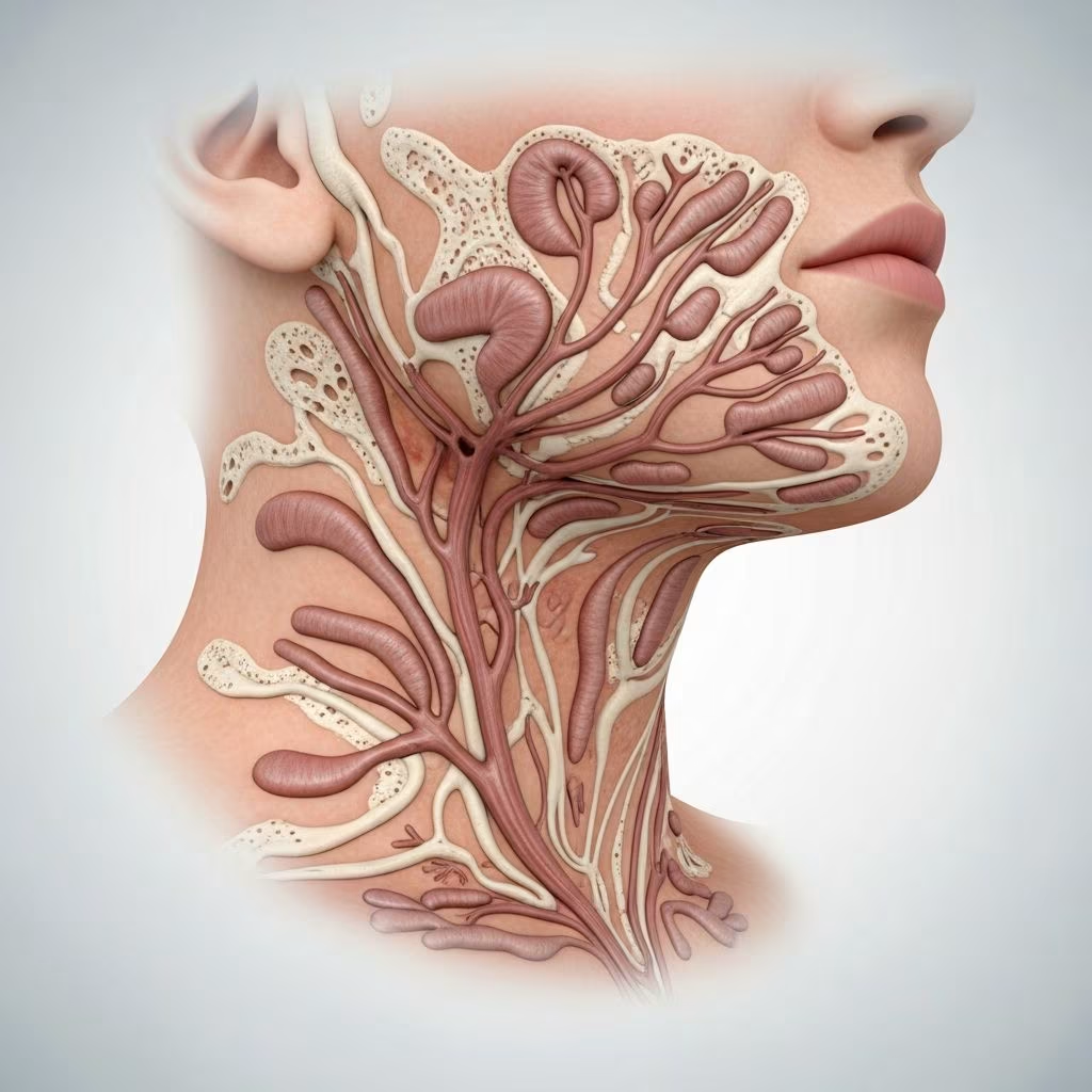
A fistula is an abnormal channel connecting two cavities or epithelialized surfaces, often draining fluids like saliva, pus, or mucus. A sinus, by contrast, is a blind-ended tract with a single external opening. These terms are frequently used interchangeably in clinical practice due to overlapping presentations.
Fistulas and sinuses in the neck and face arise from diverse etiologies, broadly classified as developmental (congenital), infectious/inflammatory, traumatic, or neoplastic. They manifest as persistent drainage, recurrent infections, swelling, or painless pits/dimples. Early recognition prevents complications like deep abscesses or intracranial spread.
What are the issues?
Patients typically present with:
- Chronic or intermittent discharge (purulent, mucoid, or serous) from a skin pit or dimple.
- Recurrent cellulitis, abscesses, or foul-smelling drainage around the opening.
- Painless midline or lateral neck pits present since birth (suggesting congenital origin).
- Swelling or mass preceding fistula formation, especially post-infection.
Locations vary: preauricular (near ear), postauricular, midline neck (thyroglossal), anterior neck triangle (branchial cleft type II), or facial (dental, nasal).
Who gets fistulas and sinuses of the neck and face?
Congenital forms appear at birth or early childhood, more common in males for branchial cleft anomalies. Acquired fistulas affect all ages, with infectious types prevalent in adults due to dental infections, trauma, or immunosuppression. Second branchial cleft cysts account for 90% of congenital neck masses.
What causes fistulas and sinuses of the neck and face?
Classified by etiology:
Developmental (congenital) causes
Arise from incomplete regression of embryonic structures:
- Branchial cleft fistulas/sinuses: From persistent branchial pouches/clefts. Type I: preauricular or parotid region. Type II (most common): anterior sternocleidomastoid, tract to tonsillar fossa. Type III: pierces carotid bifurcation. Type IV: pharyngeal pouch remnants.
- Thyroglossal duct sinuses: Midline neck, from hyoid to thyroid/foramen cecum. Move with swallowing.
- First branchial cleft anomalies: Near external auditory canal or angle of mandible, draining mucoid fluid.
- Preauricular sinuses: Common, 1-10% prevalence, pit anterior to helix.
- Nasal dermoid sinuses: Midline dorsal nose, may contain hairs, risk of intracranial extension.
- Frontocutaneous fistulas: From frontal sinus mucoceles or chronic sinusitis eroding bone.
Infectious/inflammatory causes
- Dental origin (oral-cutaneous fistulas): Most common acquired type; periapical abscess from mandibular/ maxillary teeth drains to chin, cheek, or submandibular area.
- Other infections: Staphylococcal/candidal abscesses, actinomycosis, tuberculosis, or hidradenitis suppurativa.
- Frontal sinusitis/Pott’s puffy tumor: Leads to osteomyelitis and skin fistula.
Traumatic causes
- Prior surgery, penetrating injuries, or radiation necrosis creating epithelialized tracts.
Neoplastic/malignant causes
- Rare: Squamous cell carcinoma, salivary gland tumors eroding skin.
Clinical features of specific entities
| Type | Location | Features | Tract Destination |
|---|---|---|---|
| Preauricular sinus | Anterior helix | Pit ± infection | External ear canal |
| Second branchial fistula | Anterior sternocleidomastoid | Mucoid discharge | Tonsillar fossa |
| Thyroglossal sinus | Midline neck | Moves with swallowing | Foramen cecum |
| Dental fistula | Chin/cheek | Purulent post-abscess | Periapical tooth |
| Nasal glioma/sinus | Dorsal nose | Hairs, infections | Skull base ± |
Nasal dermoid sinus tract: Distinctive dimple at nasofrontal junction with emanating hairs. Represents sequestered ectodermal cells; may extend intracranially, causing recurrent meningitis.
Diagnosis
- Clinical examination: Identify pit/opening, elicit discharge by pressure, note relation to landmarks (hyoid, mandible).
- Imaging:
- Ultrasound: Initial for cysts/sinuses.
- CT/MRI: Map tract depth, intracranial extension, bony erosion.
- Fistulogram: Contrast injection to delineate tract (less common).
- Biopsy/FNA: Rule out malignancy or confirm infection.
- Dental X-rays/panoramic for oral-cutaneous fistulas.
Treatment
Depends on etiology and symptoms:
- Asymptomatic congenital sinuses: Observation.
- Infected: Antibiotics ± incision/drainage.
- Definitive surgery:
- Stepladder incision with core-out excision of tract.
- Thyroglossal: Sistrunk procedure (central hyoid resection).
- Branchial: Complete tract excision ± fistula probe.
- Dental: Root canal/apicectomy/extraction.
- Frontocutaneous: Endoscopic sinus surgery, debridement, possible craniotomy.
Recurrence risk high if incomplete excision. Multidisciplinary approach for complex cases.
Frequently Asked Questions (FAQs)
Q: Are neck pits always congenital fistulas?
A: No, while midline pits often are, lateral pits or those with pus suggest infectious causes like dental abscesses.
Q: Can these lead to serious complications?
A: Yes, nasal sinuses risk meningitis; branchial cysts can form deep neck abscesses; frontal fistulas may cause cavernous sinus thrombosis.
Q: Is surgery always required?
A: Only for symptomatic/recurrent cases; asymptomatic preauricular sinuses may be monitored.
Q: How to differentiate dental fistula from others?
A: Proximity to jawline, dental pain history, and panoramic X-ray confirming periapical pathology.
Q: What is the prognosis after excision?
A: Excellent with complete removal; recurrence rates 10-20% for branchial clefts if hyoid not addressed in thyroglossal cases.
Related topics
- Branchial cleft cysts
- Thyroglossal duct cyst
- Dental abscess
- Pott puffy tumour
- Salivary gland infections
References
- Fistulas and Sinuses of the Neck and Face — DermNet NZ. 2023-05-15. https://dermnetnz.org/topics/fistulas-and-sinuses-of-the-neck-and-face
- Frontocutaneous Fistula — EyeWiki (American Academy of Ophthalmology). 2024-08-20. https://eyewiki.org/Frontocutaneous_Fistula
- Fistula (Neck or Face) — Ento Key. 2022-11-10. https://entokey.com/fistula-neck-or-face/
- Oral Cutaneous Fistula — MD Searchlight. 2024-03-12. https://mdsearchlight.com/oral-health/oral-cutaneous-fistula/
- Oral Cutaneous Fistula — StatPearls (NCBI Bookshelf, NIH). 2023-07-17. https://www.ncbi.nlm.nih.gov/books/NBK539822/
- Branchial Cleft Abnormalities — Children’s Hospital Colorado (.org health info). 2024-01-05. https://www.childrenscolorado.org/conditions-and-advice/conditions-and-symptoms/conditions/branchial-cleft-abnormalities/