
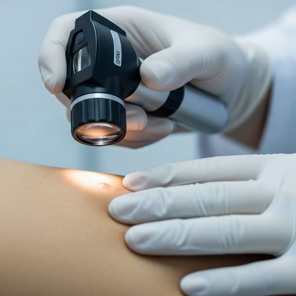
The skin, as the body’s largest organ, serves as a critical indicator of overall health. A systematic examination of the skin is essential for dermatologists and primary care physicians to detect conditions ranging from benign lesions to life-threatening skin cancers early. This process involves thorough inspection, palpation, and documentation to ensure no area is overlooked, facilitating timely intervention and improved patient outcomes.
Importance of Skin Examination
Regular skin examinations are vital for early detection of dermatological diseases. Full-body skin checks enable the identification of precancerous and cancerous lesions, with studies emphasizing annual exams for at-risk populations. Primary care physicians play a key role by performing systematic assessments, using tools like the ABCDE rule for melanoma screening. Self-exams are also recommended by the American Academy of Dermatology (AAD) to empower patients in monitoring changes.
Neglecting areas like the scalp, nails, mucous membranes, and interdigital spaces can miss critical signs. Comprehensive exams reduce mortality from skin cancer through prompt biopsy and referral.
Preparation for Skin Examination
Proper preparation ensures an effective exam. Explain the procedure to the patient, obtain consent, and provide a gown for privacy. Gather a medical history, including family history of skin cancer, sun exposure, and prior conditions.
- Ensure adequate lighting and a well-lit room.
- Prepare equipment: magnifying glass, dermatoscope, ruler, gloves, and documentation tools.
- Position the patient systematically, starting fully clothed for history, then undressed.
A chaperone should be present for sensitive exams, maintaining patient comfort.
Examination Techniques
Skin exams rely on core techniques: inspection, palpation, and specialized tests.
Inspection
Visually survey the entire body for lesions, using natural and artificial light. Note distribution, morphology, and changes. Examine front, back, sides, arms raised, and use mirrors for hard-to-see areas.
Palpation
Assess texture, consistency (soft, firm, indurated), temperature, mobility, and tenderness. Use gloved hands to feel lesions, distinguishing fluid-filled from solid.
Dermoscopy
This non-invasive tool magnifies subsurface structures, aiding diagnosis of melanoma and other lesions. Apply alcohol or gel for immersion.
Auxiliary Tests
- Biopsy: Gold standard for confirmation, including punch, shave, or excisional.
- Wood’s Lamp: Detects pigmentation and infections.
- Laboratory Studies: Cultures, scrapings for mycology.
Systematic Full-Body Skin Examination
Follow a head-to-toe protocol to avoid omissions.
- Scalp: Part hair systematically, inspect for lesions, use hand mirror.
- Face: Forehead, eyes (including canthi), nose, cheeks, lips, chin. Check asymmetry, borders.
- Ears and Neck: External canal, retro-auricular, anterior/posterior neck.
- Upper Extremities: Arms, elbows, wrists, palms, interdigital spaces, nails.
- Trunk: Chest, abdomen, back, sides; lift breasts if applicable.
- Lower Extremities: Legs, knees, ankles, feet, toes, soles.
- Genitalia and Perianal: With consent and chaperone.
- Mouth and Mucous Membranes: Lips, gums, tongue, palate.
Document with photos (zoomed in/out) and measurements. Exams typically last 10-30 minutes.
Describing Skin Lesions
Accurate description is crucial. Use: primary/secondary morphology, size, shape, borders, color, distribution, arrangement.
| Characteristic | Description | Examples |
|---|---|---|
| Morphology | Primary lesions: macule, papule, vesicle; Secondary: scale, crust | Macule (<1cm flat), Plaque (>1cm elevated) |
| Size | Measure in mm/cm; triangulate | Diameter >6mm concerning |
| Shape | Round, annular, linear | Annular (ring-like) |
| Configuration | Linear, serpiginous, herpetiform | Grouped vesicles |
| Distribution | Localized, generalized, dermatomal | Symmetrical on extremities |
ABCDE Rule for melanoma: Asymmetry, Border irregularity, Color variation, Diameter >6mm, Evolution. Ugly duckling sign: lesion differing from others.
Examination of Specific Sites
Hands
Inspect palms, dorsum, nails for erythema, pigmentation, nodes.
| Finding | Examples | Associated Disease |
|---|---|---|
| Palmar erythema | Red palms | Liver disease |
| Hyperpigmentation | Brown palms | Addison disease |
| Heberden/Bouchard nodes | Joint swellings | Osteoarthritis |
| Gottron papules | Violaceous papules | Dermatomyositis |
| Janeway/Osler lesions | Nontender/tender nodules | Infective endocarditis |
Nails
Check pitting, onycholysis, clubbing, Beau lines indicating systemic issues.
Mouth and Mucous Membranes
Examine for ulcers, white patches (leukoplakia), erythema.
Scalp
Part hair for alopecia, scaling, tumors.
Suspicious Lesions and Next Steps
Atypical features: asymmetry, irregular borders, multicolored, >6mm, evolving. Refer or biopsy suspicious lesions.
- Educate on self-exams and sun protection.
- Schedule follow-ups for monitoring.
Frequently Asked Questions (FAQs)
Q: How often should I perform a full-body skin exam?
A: Annually by a clinician, plus monthly self-exams, especially for high-risk individuals.
Q: What is dermoscopy and when is it used?
A: A magnifying tool for subsurface lesion views, ideal for pigmented lesions.
Q: Are self-skin exams effective?
A: Yes, they help detect changes early when treatable.
Q: What if I find a suspicious lesion?
A: Seek medical evaluation promptly for biopsy if needed.
Q: Does skin type affect exam frequency?
A: Fair-skinned, high-sun-exposure individuals need more frequent checks.
References
- Principles of dermatology — AMBOSS. 2023. https://www.amboss.com/us/knowledge/principles-of-dermatology/
- Dermatological guide for primary care physicians: full body skin examination — PMC (NCBI). 2024-05-15. https://pmc.ncbi.nlm.nih.gov/articles/PMC11188824/
- Skin Cancer: Screening — United States Preventive Services Taskforce (USPSTF). 2024. https://www.uspreventiveservicestaskforce.org/uspstf/recommendation/skin-cancer-screening
- The Importance of Skin Examinations: Who Needs Them and Current Guidelines — Wyoming Academy of Dermatology. 2023-10-01. https://www.wyomingacademyofdermatology.org/dermatologist-insights/the-importance-of-skin-examinations-who-needs-them-and-current-guidelines
- Approach to the Dermatology Exam — Stanford Medicine 25 (YouTube). 2015. https://www.youtube.com/watch?v=qI4E9JDs2Tw
- Your Guide to Full Body Skin Exams — Affiliated Dermatology. 2024. https://affderm.com/full-body-skin-exam-guide/
- Find skin cancer: How to perform a skin self-exam — American Academy of Dermatology (AAD). 2024. https://www.aad.org/public/diseases/skin-cancer/find/check-skin



