
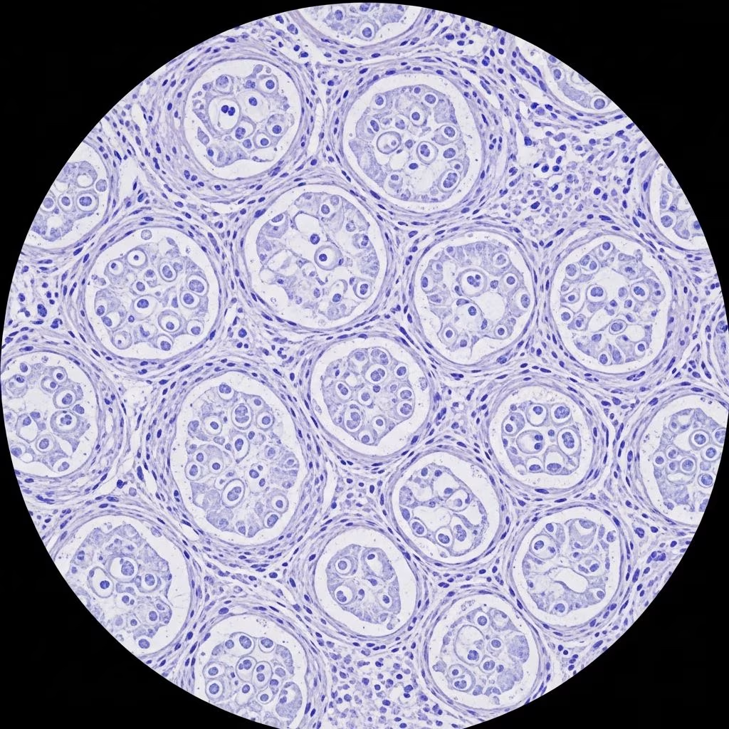
Granular cell tumours (GCTs) represent uncommon soft tissue neoplasms predominantly benign in nature, characterised by distinctive granular cytoplasm in tumour cells believed to derive from Schwann cells. These lesions pose diagnostic challenges due to their infiltrative growth and overlap with other entities, necessitating careful histopathological evaluation.
Definition / typical H&E appearance
On routine haematoxylin and eosin (H&E) staining, granular cell tumours exhibit poorly circumscribed nests and sheets of large polygonal cells infiltrating the dermis and subcutis. Tumour cells feature abundant pale eosinophilic granular cytoplasm, small eccentric nuclei with inconspicuous nucleoli, and lack significant atypia in benign cases. The granular appearance arises from lysosome accumulation, imparting a distinctive texture visible at low to high power magnification.
Intercellular collagen bundles partially separate cell clusters, contributing to the firm clinical texture. Pseudoepitheliomatous hyperplasia of the overlying epidermis is frequent, mimicking squamous cell carcinoma and complicating initial clinical assessment.
Clinical features
Clinically, GCTs manifest as solitary, firm, painless subcutaneous nodules measuring 0.5–3 cm, most prevalent on the head, neck, and oral cavity (tongue comprising up to 40% of cases). Skin involvement occurs in 30–40% of instances, with females affected twice as often as males, peaking in the 4th–6th decades.
- Skin-coloured to yellowish-brown papules or nodules
- Slow-growing, asymptomatic
- Multiple lesions in 10–25% of patients, especially those with dark skin
- Associated syndromes: neurofibromatosis type 1, LEOPARD, Noonan
Malignant features include rapid growth, size >5 cm, pain, and deep location.
Histopathology and microscopy
Cytology description
- Large epithelioid to spindle cells with ill-defined borders
- Abundant granular eosinophilic cytoplasm
- Small round/oval nuclei, even chromatin, small nucleoli
- Low mitotic activity (<2/10 HPF in benign cases)
- No necrosis or vascular invasion typically
Microscopic (histologic) images
Low-power view reveals dermal infiltrative nests amid collagen; high-power shows granular cytoplasm with pyknotic nuclei. PAS stain highlights diastase-resistant intracytoplasmic granules.
Cytology images
Cytological smears display dispersed polygonal cells with granular cytoplasm, stripped nuclei, and prominent lysosomes, aiding fine-needle aspiration diagnosis.
Electron microscopy description
Ultrastructurally, tumour cells contain numerous lysosomes, autophagosomes, and myelin-like figures, supporting Schwannian differentiation. No true myofilaments observed, refuting obsolete myoblastoma nomenclature.
Pathology molecular biology
Mutations in TSC1/TSC2 (mTOR pathway) and MAPK/ERK pathway alterations underlie GCT pathogenesis. Benign lesions show few copy number variations; malignant cases exhibit higher genomic instability.
Immunohistochemistry and special stains
| Marker | Benign GCT | Malignant GCT |
|---|---|---|
| S100 | Diffuse strong + | Positive (variable) |
| SOX10 | Strong + | Positive |
| CD68 | Focal + (lysosomes) | Focal + |
| Ki67 (<3%) | Low | |
| p53 | Low | High in malignant |
| EMA, GFAP, desmin | – | – |
S100 and SOX10 positivity confirms neural origin; CD68 reflects phagolysosomes. PAS+ diastase-resistant granules diagnostic.
Boarding / frozen section description
Intraoperative frozen sections show granular cells but IHC unavailable; recommend wide excision over partial biopsy due to infiltrative margins.
Cytology patterns
- Dispersed polygonal granular cells
- Thick colloid-like stroma
- Vacuolated cytoplasm
Differential diagnosis
| Entity | Key Distinguisher |
|---|---|
| Rhabdomyoma | Skeletal muscle IHC (myogenin+), no granules |
| Hibernoma | |
| Adult granulosa cell tumour | Groer pattern, inhibin+, ovarian |
| Leiomyoma | Smooth muscle actin+, no granules |
| Malignant GCT | Atypia, necrosis, mitoses >2/10HPF, size >5cm |
| Schwannoma | Antoni A/B, Verocay bodies, encapsulated |
Malignancy criteria (Fanburg-Smith)
Malignant GCTs (<2%) defined by ≥3 features or metastasis:
- Nucleolar pleomorphism
- High N:C ratio
- Mitoses >2/10 HPF
- Necrosis
- Spindling
- Vesicular nuclei
Atypical: 1–2 features; monitor closely.
Management and prognosis
Benign GCTs cure by complete surgical excision with 5mm margins; recurrence rare (<5%) if margins clear. Malignant cases require wide excision, lymph node dissection, and surveillance; metastasis to lymph nodes/lungs portends poor prognosis (50% mortality).
No role for adjuvant therapy routinely.
Frequently Asked Questions (FAQs)
Q: Are granular cell tumours cancerous?
A: Over 98% benign; malignancy rare, indicated by size >5cm, atypia, high mitoses.
Q: What is the origin of granular cell tumours?
Q: Schwann cells (S100/SOX10+); granular cytoplasm from lysosomes.
Q: How are GCTs treated?
A: Wide local excision; Mohs unsuitable due to poor margination.
Q: Can GCTs recur?
A: <5% benign if completely excised; malignant recur/metastasize frequently.
Q: Is IHC always positive for S100 in GCT?
A: Yes, diffuse strong; essential for diagnosis.
Related topics
- Schwannoma pathology
- Neurofibroma pathology
- Soft tissue tumours
- S100 positive markers
References
- Granular cell tumour of the skin — PRRS Journal. 2023. https://www.prrsjournal.com/article/view/189
- An update on cutaneous granular cell tumours for dermatologists — PubMed / British Journal of Dermatology. 2022-06-01. https://pubmed.ncbi.nlm.nih.gov/35727729/
- Granular Cell Tumours — DermNet NZ (official dermatology resource). 2023. https://dermnetnz.org/topics/granular-cell-tumour



