
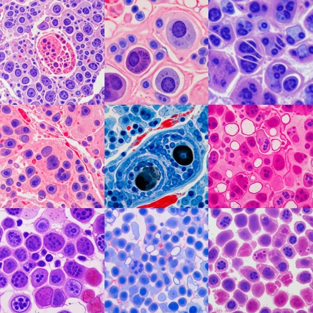
Histology stains are fundamental tools in dermatopathology, enabling pathologists to visualize tissue structures, identify cellular components, and diagnose skin diseases. Routine staining with haematoxylin and eosin (H&E) provides a baseline view, while special stains and immunohistochemistry target specific substances for detailed analysis.
What are histology stains?
Histological staining involves applying dyes to tissue sections to highlight specific structures under a microscope. In dermatopathology, these stains differentiate normal from pathological skin components, such as inflammation, neoplasms, or infections. Haematoxylin, being basic, stains acidic structures blue-purple, while eosin, acidic, stains basic structures pink-red. Special stains selectively bind to molecules like fungi, melanin, or amyloid, supplementing H&E when routine views are insufficient.
These techniques are crucial for diagnosing conditions like dermatitis herpetiformis, melanoma, or fungal infections, where H&E alone may not suffice. Immunohistochemistry (IHC) further enhances specificity using antibodies against cellular markers.
Common histological stains
The following table lists common stains used in dermatopathology, their typical colors, and primary applications.
| Stain | Color | Typical use |
|---|---|---|
| Bielschowsky stain | Black | Neural plaques and tangles |
| Congo red | Red | Amyloid fibres |
| Crystal violet | Violet | Glia and neurons |
| Eosin | Pink/orange/red | General staining with haematoxylin (cytoplasm, collagen) |
| Fontana-Masson | Black/pink or red | Melanin |
| Giemsa | Blue/violet/pink | Blood/bone marrow smears, bacteria, protozoa, mast cells |
| Haematoxylin | Blue/purple | General staining with eosin (nuclei) |
| Luna stain | Purple/black | Mast cells and elastin |
| Nissl | Blue | Rough endoplasmic reticulum in neurons |
| Periodic acid-Schiff (PAS) | Red/magenta | Glycogen, basement membranes, fungi, mucins |
| Oil Red O | Red | Fat emboli, lipids |
Routine stains
Haematoxylin and eosin (H&E)
The cornerstone of dermatopathology, H&E is the first stain applied to skin biopsies. Haematoxylin stains basophilic structures like nuclei and the epidermal granular layer blue-purple. Eosin stains eosinophilic elements such as cytoplasm, collagen, muscle, and fibrin pink-red. This predictable pattern elucidates basic histological features, including architecture, inflammation, and preliminary microorganism detection.
Uses include assessing calcification, fungi, bacteria, and as a precursor to special or immunohistochemical studies. In skin biopsies, it reveals epidermal thickness (7-12 cell layers), dermal structures, and adipose layers. For example, in dermatitis herpetiformis, H&E shows neutrophils in dermal papillae and subepidermal vesicles.
Special stains
Special stains complement H&E by targeting specific tissue components, such as microorganisms, connective tissues, or pigments. They are essential in inflammatory and neoplastic skin diseases where routine staining falls short.
Gram stain
Gram stain differentiates bacteria: Gram-positive appear purple, Gram-negative pink. Vital for identifying bacterial infections in skin biopsies.
Gomori methenamine silver (GMS)
GMS stains fungal cell walls and parasites brown-black against a green background. Commonly used for Pneumocystis carinii, Histoplasma spp., and Leishmania. It highlights fungi not visible on H&E.
Giemsa stain
This stain colors bacteria blue, cytoplasm pink-rose, nuclei blue, and mast cells purple. Ideal for bacteria, protozoa, and mast cell evaluation in dermatological specimens.
Periodic acid-Schiff (PAS)
PAS produces red-magenta staining for glycogen, basement membranes, reticular fibres, cartilage, glycoproteins, glycolipids, and mucins. Excellent for fungal infections like candidiasis and basement membrane thickening in lichen planus.
Fontana-Masson
Stains melanin black, pink, or red, aiding diagnosis of melanocytic lesions like melanoma.
Warthin-Starry
Detects spirochetes like those in syphilis, staining them black.
Other special stains include Van Gieson for collagen and smooth muscle differentiation, Luna for mast cells and elastin, and Congo red for amyloid, which shows apple-green birefringence under polarized light.
Immunohistochemistry (IHC) stains
IHC uses antigen-antibody binding for precise identification of cells and substances, often replacing older histochemical methods. Antibodies target cytoskeletal proteins, melanocytic markers, or lymphoid differentiation.
| Antibody Target | Antibody | Identifies |
|---|---|---|
| Intermediate Filaments | Cytokeratin | Epithelial differentiation |
| Intermediate Filaments | Desmin | Muscle differentiation |
| Intermediate Filaments | Actin | Muscle (less specific) |
| Melanocytic differentiation | S100 | Sensitive, non-specific |
| Melanocytic differentiation | HMB-45 | More specific, less sensitive |
| Melanocytic differentiation | Melan-A | Specific and sensitive |
In immunoperoxidase IHC, peroxidase enzymes produce brown staining; fluorescent IHC emits under microscopy. Common in melanoma (S-100, SOX10, MART-1, HMB-45) and lymphomas[10]. Characteristic patterns aid immunobullous diseases, lupus, and vasculitis.
Other tests
Beyond stains, direct immunofluorescence detects antibodies in biopsies using fluorescein-labelled probes, revealing patterns in pemphigus or bullous pemphigoid. Indirect immunofluorescence uses patient serum on substrates for circulating antibodies. Fluorescent in situ hybridization (FISH) identifies chromosomal changes in cutaneous lymphomas.
Frequently Asked Questions (FAQs)
What is the primary use of H&E stain in dermatopathology?
H&E provides a standard view of tissue architecture, staining nuclei blue-purple and cytoplasm/collagen pink-red, essential for initial biopsy assessment.
When are special stains necessary?
Special stains are used when H&E fails to identify specific pathogens, pigments, or substances like fungi (GMS, PAS) or melanin (Fontana-Masson).
How does IHC differ from special stains?
IHC uses specific antibodies for targeted protein detection, offering higher specificity than histochemical special stains.
Which stain detects amyloid in skin biopsies?
Congo red stains amyloid red, with apple-green birefringence under polarized light.
Can histology stains confirm melanoma?
Fontana-Masson stains melanin, while IHC markers like S100 and HMB-45 confirm melanocytic origin[10].
These stains and techniques ensure accurate dermatopathological diagnoses, guiding clinical management from infections to malignancies. Advances in IHC and molecular tests continue to refine precision.
References
- Stains in Dermatology — Scribd. 2022-10-01. https://www.scribd.com/presentation/735058382/Stains-in-Dermatology
- Histology stains – DermNet — DermNet NZ. 2023-01-15. https://dermnetnz.org/topics/histology-stains
- Dermatopathology. Special stains and tests – DermNet — DermNet NZ. 2024-05-20. https://dermnetnz.org/cme/dermatopathology/special-stains-and-tests
- What is dermatopathology? – DermNet — DermNet NZ. 2023-11-10. https://dermnetnz.org/topics/dermatopathology
- Introduction to dermatopathology – DermNet — DermNet NZ. 2024-02-28. https://dermnetnz.org/topics/introduction-to-dermatopathology



