
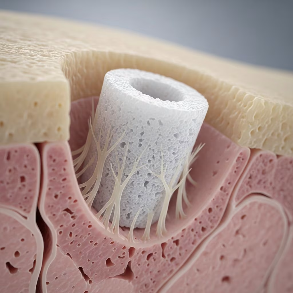
Hydroxyapatite (HA) implants are biocompatible materials mimicking the mineral composition of human bone, widely used in medical fields for bone regeneration, implants, and prosthetics due to their excellent integration with body tissues.
What is hydroxyapatite?
Hydroxyapatite, chemically Ca10(PO4)6(OH)2, constitutes approximately 65-70% of human bone and 97% of tooth enamel, providing structural integrity and hardness. Synthetically produced HA is engineered to replicate this natural form, offering osteoconductive properties that guide bone growth along its surface and bioactive qualities that stimulate biological responses for healing.
In implants, HA’s biocompatibility ensures the immune system does not reject it as foreign, facilitating seamless integration with surrounding tissues. Its stability, low solubility, and ability to interact positively with osteoblasts—bone-forming cells—make it superior for long-term applications. Unlike resorbable materials, dense HA maintains structural support while promoting natural bone apposition.
Who gets a hydroxyapatite implant and why?
Hydroxyapatite implants are indicated for patients requiring bone augmentation or defect repair, particularly in dermatological reconstruction, orthopedics, dentistry, and maxillofacial surgery. Common scenarios include:
- Facial contouring and defect filling in dermatologic or reconstructive procedures post-trauma, tumor resection, or congenital deformities.
- Dental implant support where bone volume is insufficient, such as ridge augmentation or sinus lifts.
- Orthopedic applications like hip/knee replacements, spinal fusions, and fracture repairs needing scaffolds for bone regrowth.
The primary rationale is HA’s ability to promote osseointegration—direct bone-to-implant contact—enhancing stability and reducing failure rates. It serves as an alternative to autografts, avoiding donor site morbidity while providing a scaffold for new bone formation.
Clinical features of hydroxyapatite implants
HA implants appear as white, porous or dense ceramic-like structures, customizable in forms like blocks, granules, powders, or coatings. Post-implantation, initial bioactivity triggers ion exchange with body fluids, forming a carbonated apatite layer that bonds with bone.
| Property | Description | Benefit |
|---|---|---|
| Biocompatibility | Structurally identical to bone mineral | Minimal rejection risk |
| Osteoconductivity | Supports bone growth on surface | Faster healing |
| Bioactivity | Stimulates tissue responses | Enhanced integration |
| Forms | Coatings, scaffolds, granules | Versatile applications |
Clinically, successful implants show radiographic bone density increase over 6-12 months, with no inflammation.
Histology of hydroxyapatite-tissue interface
Microscopically, HA interfaces exhibit direct appositional bone formation without fibrous encapsulation, unlike non-bioactive metals. Osteoblasts attach to HA’s textured surface, proliferating and depositing matrix that mineralizes into new bone. Scanning electron microscopy reveals intimate bone-HA contact, with gradual replacement by host bone in porous variants. This osteointegration is mediated by HA’s calcium phosphate release, mimicking natural remineralization. Studies confirm increased osteoblast activity and reduced osteoclast resorption, ensuring long-term stability.
Types of hydroxyapatite implants
- Dense HA: High crystallinity for load-bearing, used in orbital floor implants or facial prostheses; slow resorption.
- Porous HA: Interconnected pores (200-500μm) allow vascular ingrowth and faster bone infiltration; ideal for large defects.
- Granular/Particulate HA: For bone void fillers in grafting; injectable forms for minimally invasive use.
- HA Coatings: Plasma-sprayed onto titanium implants (50-200μm thick) for dental/orthopedic use, enhancing early fixation.
- Nano-HA: Smaller particles for superior bioactivity in enamel repair or thin coatings.
Indications for hydroxyapatite implants
Key indications include:
- Periodontal defects: Intrabony fills for regeneration.
- Alveolar ridge augmentation: Pre-dental implant bone building.
- Sinus lifts: Maxillary floor elevation.
- Orthopedic: Joint replacements, spinal fusions, non-union fractures.
- Maxillofacial: Cranial/facial reconstructions.
- Dermatologic: Subdermal fillers for contouring scars or atrophic skin.
Hydroxyapatite implant procedure
The procedure involves:
- Pre-op planning: Imaging (CBCT) assesses defect; HA selected based on site/load.
- Anesthesia: Local/general; flap elevation exposes defect.
- Preparation: Decortication for bleeding bone bed.
- Implantation: HA placed/condensed; coated implants screwed in.
- Stabilization: Membranes/collagen barriers for guided regeneration; closure.
- Post-op: Antibiotics, soft diet; follow-up at 1-2 weeks.
Integration occurs in phases: inflammatory (days), reparative (weeks), remodeling (months).
Alternatives to hydroxyapatite
| Material | Pros | Cons vs HA |
|---|---|---|
| Autograft | Gold standard, osteoinductive | Donor morbidity, limited volume |
| Allograft/Xenograft | Readily available | Disease risk, slower integration |
| Beta-TCP | Resorbable | Weaker mechanically |
| Bioactive Glass | Fast bonding | Brittle, variable resorption |
| Titanium (uncoated) | Strong | No bioactivity, fibrous tissue |
HA excels in non-resorbable, stable support.
Outcomes and complications
Success rates exceed 90% for dental osseointegration, with 5-year survival >95%. Orthopedic coatings reduce loosening by 30-50%. Complications (rare, <5%):
- Early: Infection, exposure, mobility.
- Late: Non-integration, fracture (dense HA brittle).
- Mitigation: Proper case selection, sterile technique.
Prevention of complications
- Aseptic surgery, prophylactic antibiotics.
- Adequate defect vascularity.
- Overcorrection for remodeling.
- Regular radiographic monitoring.
References in context
HA’s efficacy is backed by decades of use, from bone grafts over a century ago to modern coatings.
Frequently Asked Questions
What is a hydroxyapatite implant used for?
Used for bone regeneration in dental, orthopedic, and reconstructive surgeries by providing a scaffold that promotes natural bone growth.
Is hydroxyapatite safe for implants?
Yes, highly biocompatible with low rejection risk due to its natural bone-like composition.
How long does hydroxyapatite implant integration take?
Initial stability in weeks; full osseointegration in 3-6 months, depending on site.
Can hydroxyapatite implants be resorbed?
Dense forms are non-resorbable; porous/granular types partially replaced by bone over time.
What are the advantages over other bone substitutes?
Superior osteoconductivity, no disease transmission, and mechanical stability.
References
- What is Hydroxyapatite? — Supermouth. 2023. https://supermouth.com/learn/what-is-hydroxyapatite
- How Is Hydroxyapatite Used In Implants? — YouTube (Video Transcript). 2023. https://www.youtube.com/watch?v=73iqrYgsO-4
- Hydroxyapatite for Dental Care — Arbor Falls Dental. 2024. https://arborfallsdentalandwellness.com/service/hydroxyapatite-jackson-mi/
- Hydroxyapatite and its biomedical application — Biovac. 2023. https://biovac.es/en/hydroxylapatite-and-its-biomedical-application/
- Benefits of hydroxyapatite coatings on medical implants — HiMed. 2023. https://www.himed.com/blog/benefits-hydroxyapatite-coatings-ortho-dental-implants
Disclaimer: This information is for educational purposes only and not a substitute for professional medical advice. Consult a healthcare provider for personalized guidance.



