
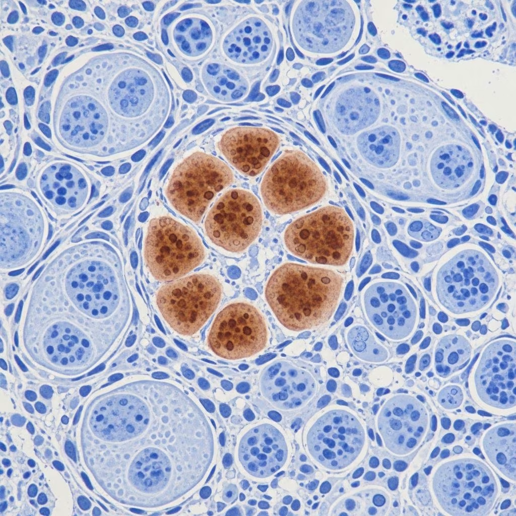
Immunohistochemistry (IHC) is a powerful laboratory technique that uses antibodies to detect specific antigens in tissue sections, enabling precise identification of cell types, tumours, and pathological processes in dermatopathology. Hundreds of IHC stains are employed to differentiate neoplasms, with a select few commonly used in dermatology for enhanced diagnostic accuracy beyond standard haematoxylin and eosin (H&E) staining.
What is immunohistochemistry?
Immunohistochemistry involves applying primary antibodies that bind to target proteins (antigens) in fixed tissue samples. A secondary antibody, often linked to an enzyme like peroxidase or a fluorescent tag, amplifies the signal, producing a visible stain—typically brown in immunoperoxidase methods or fluorescence under a microscope. This method bridges histology and molecular biology, revealing protein expression, localisation, and cellular interactions critical for diagnosing skin conditions.
In dermatology, IHC is indispensable for distinguishing poorly differentiated tumours, confirming margins in Mohs micrographic surgery (MMS), and identifying infectious agents or prognostic markers. It supports personalised medicine by detecting biomarkers like HER2 for targeted therapies.
How does IHC staining work?
The IHC process begins with tissue fixation, typically in formalin, followed by paraffin embedding and sectioning. Antigen retrieval unmasks epitopes hidden by fixation. Primary antibodies (polyclonal for sensitivity or monoclonal for specificity) bind antigens, followed by secondary enzyme-conjugated antibodies. Substrates like DAB (diaminobenzidine) produce chromogenic precipitates, while counterstains like haematoxylin highlight nuclei.
Fluorescent IHC uses fluorochromes for multiplex detection, allowing simultaneous visualisation of multiple markers. Tyramide signal amplification (TSA) enhances weak signals. Single IHC targets one antigen; multiplex IHC analyses several, revealing tissue heterogeneity and signalling pathways.
Common IHC stains in dermatology
Dermatopathology relies on targeted IHC panels. Below is a table summarising key stains, their targets, applications, and limitations, drawn from clinical use in skin neoplasms.
| Stain | Target | Primary Use | Strengths | Limitations |
|---|---|---|---|---|
| MART-1 (Melan-A) | Melanocyte protein | Melanoma in situ, invasive melanoma | High sensitivity for melanocytes; 1-hour protocol in MMS | Stains benign nevi; poor for spindle/desmoplastic melanoma |
| SOX10 | Nuclear transcription factor in melanocytes/neural crest | Desmoplastic/spindle cell melanoma | Superior sensitivity for neurotropic melanoma | Stains benign neural elements, scar tissue |
| PanCK (Pan-cytokeratin) | Epithelial cytokeratins | Poorly differentiated squamous cell carcinoma (SCC) | Highlights subtle epithelial tumour cells | May stain benign epithelium like sebaceous glands |
| Ber-EP4 | Epithelial cell adhesion molecule (EpCAM) | Basal cell carcinoma (BCC), basosquamous tumours | Differentiates BCC from follicles/superficial BCC | Less specific in mixed tumours |
| CD34 | Endothelial/pericytic marker | Dermatofibrosarcoma protuberans (DFSP) | Distinguishes DFSP from scar/dermatofibroma | Stains vessels, fibroblasts |
| CK7 (Cytokeratin 7) | Glandular cytokeratins | Extramammary Paget disease (EMPD) | Detects pagetoid spread | Overlaps with metastatic adenocarcinoma |
| S100 | Calcium-binding protein | Melanocytic, neural tumours | Broad sensitivity for neural/melanocytic lesions | Non-specific; stains Langerhans cells, sweat glands |
| Factor XIIIa | Dermal dendrocyte marker | Dermatofibroma vs. DFSP | Highlights dendritic cells in benign fibrous tumours | Variable in atypical lesions |
Applications in skin tumours
Melanoma
MART-1 and SOX10 are staples in MMS for melanoma. MART-1 stains cytoplasmic melanocyte markers effectively in conventional melanoma but misses desmoplastic variants. SOX10, targeting nuclear antigens, excels in neurotropic and spindle cell types, aiding margin clearance despite potential false positives from neural scars.
Basal cell carcinoma
Ber-EP4 differentiates BCC from benign adnexal structures, crucial in superficial or morpheaform BCC where H&E is ambiguous. It highlights basosquamous differentiation, guiding surgical excision.
Squamous cell carcinoma
PanCK is vital for poorly differentiated SCC, where tumour cells mimic inflammation. In MMS, it confirms residual disease, preventing over- or under-treatment.
Other neoplasms
CD34 delineates DFSP infiltrative tendrils from fibrosis. CK7 identifies EMPD pagetoid cells, distinguishing primary from secondary disease. S100 aids melanocytic/Schwannian tumours, while Ki-67 assesses proliferation.
Interpretation of IHC results
IHC patterns guide diagnosis:
- Nuclear staining: Transcription factors (e.g., SOX10, p53, Ki-67). Indicates proliferative or regulatory activity.
- Cytoplasmic staining: Structural/enzymatic proteins (e.g., cytokeratins, MART-1). Suggests cell lineage.
- Membranous staining: Receptors/adhesion molecules (e.g., Ber-EP4, HER2). Relevant for targeted therapies.
Quantify intensity (0-3+), distribution (focal/diffuse), and controls. Correlate with H&E; discordant results warrant repeat staining or additional markers.
Advantages and limitations
Advantages:
- Enhances precision in challenging cases, reducing recurrence in MMS.
- Supports prognosis (e.g., Ki-67) and therapy selection.
- Multiplexing reveals tumour microenvironment.
Limitations:
- Antibody specificity/sensitivity varies; cross-reactivity possible.
- Fixation artefacts, antigen retrieval inconsistencies affect results.
- Costly, time-intensive for frozen MMS sections.
- Interpretation requires expertise; over-reliance risks misdiagnosis.
IHC in Mohs surgery
In MMS, rapid IHC protocols (e.g., 1-hour MART-1) facilitate real-time margin assessment for melanoma, SCC, and adnexal tumours. PanCK clarifies ambiguous SCC, SOX10 unmasks desmoplastic melanoma, improving clearance rates and conserving tissue.
Frequently asked questions
What is the difference between H&E and IHC staining?
H&E provides general morphology; IHC adds molecular specificity via antigen detection, essential for tumour subtyping.
Which IHC stain is best for melanoma in Mohs?
SOX10 for desmoplastic types; MART-1 for conventional. Use panels for accuracy.
Can IHC distinguish benign from malignant lesions?
Partially; e.g., CD34+ honeycomb in DFSP vs. patchy in dermatofibroma. Combine with histology.
How long does IHC take in clinical practice?
Standard: 4-24 hours; rapid MMS protocols: 1-2 hours.
Is IHC useful for infectious diseases?
Yes, detects pathogens like Treponema or viruses via specific antibodies.
References
- The Role of Immunohistochemical Stains in Mohs Surgery — Dermatology Times. 2023. https://www.dermatologytimes.com/view/the-role-of-immunohistochemical-stains-in-mohs-surgery
- Immunohistochemistry stains – DermNet — DermNet NZ. 2024. https://dermnetnz.org/topics/immunohistochemistry-stains
- Immunohistochemistry (IHC) staining: Techniques and applications — Abcam. 2024. https://www.abcam.com/en-us/knowledge-center/immunohistochemistry/ihc-staining
- IMMUNOHISTOCHEMISTRY: RELEVANCE IN DERMATOLOGY — NIH/PMC. 2012-01-01. https://pmc.ncbi.nlm.nih.gov/articles/PMC3276886/
- Useful Immunohistochemical Stains for Differentiating Various Skin Conditions — Journal of Drugs in Dermatology. 2014-05-01. https://jddonline.com/articles/resident-rounds-part-ii-useful-immunohistochemical-stains-for-differentiating-various-skin-condition-S1545961614P0613X



