
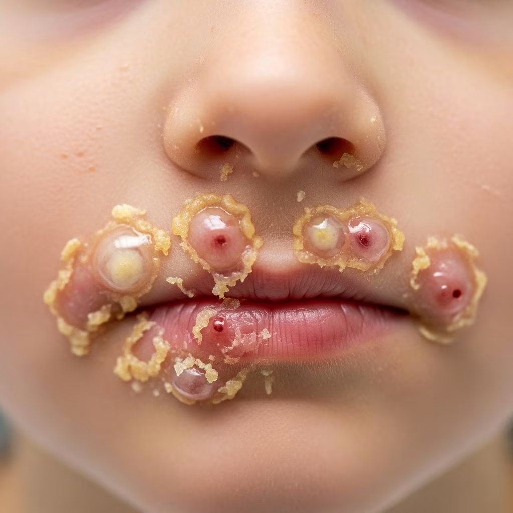
What is impetigo?
Impetigo is a common and highly contagious bacterial skin infection that primarily affects the superficial layers of the epidermis. It is most prevalent in children, particularly those aged 2–5 years, but can occur in people of any age. The infection typically presents as erythematous plaques topped with characteristic yellow or honey-coloured crusts, which may be itchy or painful. Impetigo spreads easily through direct contact or shared items, thriving in warm, humid environments and close-contact settings like schools or daycare centres.
Without treatment, impetigo usually resolves spontaneously within 2–3 weeks, but antibiotic therapy accelerates healing to about 7–10 days and reduces transmission risk. Although generally mild, untreated cases can lead to complications such as deeper infections or, rarely, post-streptococcal glomerulonephritis.
Who gets impetigo?
Impetigo predominantly affects young children, with peak incidence in preschool and school-aged groups. Risk factors include:
- Warm, humid climates
- Crowded living conditions
- Poor hygiene
- Pre-existing skin trauma (cuts, abrasions, insect bites)
- Underlying skin conditions like atopic dermatitis or scabies
- Immunosuppression or diabetes
Adults are less commonly affected but may be at higher risk for complications if infected. Nasal carriers of Staphylococcus aureus are prone to recurrent episodes.
Causes
Impetigo is caused by gram-positive bacteria, primarily Staphylococcus aureus (including MRSA strains) and Streptococcus pyogenes (group A streptococcus). These pathogens invade through minor skin breaks. Secondary impetigo often complicates existing dermatoses.
Types of impetigo
Two main clinical forms exist: non-bullous (most common, 70% of cases) and bullous impetigo.
Non-bullous impetigo
Accounts for the majority of cases. Begins as small vesicles or pustules on erythematous skin that rupture, releasing seropurulent fluid which dries into variably sized, golden-yellow crusts. Lesions are typically on exposed areas like the face (perioral, perinasal), extremities. Regional lymphadenopathy may occur; fever is rare.
Bullous impetigo
Caused exclusively by exfoliative toxin-producing S. aureus. Features flaccid, transparent bullae (1–2 cm) filled with clear yellow fluid that progresses to pus. Bullae rupture leaving a collarette of scale. Prefers trunk, axillae, buttocks, diaper areas. Less contagious than non-bullous form.
Clinical features
Nonbullous impetigo
Lesions evolve rapidly: papules → vesicles/pustules → erosions with honey-coloured crusts on erythematous base. Itching prompts scratching, spreading infection via autoinoculation. Common sites: nose, mouth, limbs. Incubation: 4–10 days.
Bullous impetigo
Small vesicles enlarge to flaccid bullae without significant surrounding erythema. Fluid becomes cloudy; ruptures form superficial erosions with lacelike rim. Mildly symptomatic.
Lesion morphology
| Type | Primary Lesion | Evolution | Common Sites |
|---|---|---|---|
| Non-bullous | Vesicle/pustule | Ruptures → honey crust | Face, extremities |
| Bullous | Flaccid bulla | Ruptures → erosions, scale rim | Trunk, diaper area |
Diagnosis
Clinical diagnosis based on characteristic morphology. No routine testing needed for uncomplicated cases. Bacterial culture/swab if treatment failure, recurrent infection, or high-risk patients (neonates, immunosuppressed). Gram stain shows gram-positive cocci in clusters (S. aureus) or chains (S. pyogenes). Rarely, biopsy for atypical presentations.
Differential diagnosis
- Herpes simplex
- Varicella zoster
- Contact dermatitis
- Atopic dermatitis (infected)
- Scabies
- Candidiasis
- Tinea corporis
- Pediculosis
Key differentiators: impetigo crusts are golden-yellow and ‘stuck-on’ appearance; rapid spread.
Complications
Most cases heal without sequelae. Potential issues include:
- Cellulitis: Extension to dermis
- Ecthyma: Ulcerative form with scarring
- Lymphangitis/adenitis
- Post-streptococcal glomerulonephritis (1–2%, 2–3 weeks post-infection)
- Sepsis (rare, high-risk patients)
- Pigmentary changes (hypo/hyperpigmentation)
Scarring uncommon except ecthyma.
Impetigo treatment
General measures
- Gentle cleansing with soap/water to remove crusts
- Avoid scratching
- Cover lesions
- Exclude from school/daycare until crusts dry (24–48h antibiotics)
Topical antibiotics (limited disease)
| Agent | Dosage | Duration |
|---|---|---|
| Mupirocin 2% ointment | TID | 5–7 days |
| Fusidic acid ointment | TID | 7–10 days |
| Retapamulin 1% ointment | BID | 5 days |
Fusidic acid avoided in MRSA-prevalent areas.
Oral antibiotics (widespread, bullous, high-risk)
| Agent | Adult Dose | Child Dose | Duration |
|---|---|---|---|
| Cephalexin | 500mg QID | 25–50mg/kg/d divided QID | 7 days |
| Dicloxacillin | 500mg QID | 25mg/kg/d divided QID | 7 days |
| Erythromycin | 500mg QID | 40mg/kg/d divided QID | 7 days |
| Clindamycin (MRSA) | 300–450mg TID | 20mg/kg/d divided TID | 7 days |
Anti-streptococcal coverage if GAS suspected.
Neonatal/severe cases
IV vancomycin/ceftriaxone ± clindamycin pending cultures.
Non-antibiotic options
Hydrogen peroxide 1% cream (UK): TID for 5 days. Less effective than antibiotics.
Prevention of impetigo
- Hand hygiene
- Avoid nose-picking (S. aureus reservoir)
- Cover abrasions promptly
- Daily chlorhexidine washes in outbreaks
- Mupirocin nasal decolonization for recurrent cases/carriers
- Exclude cases until non-infectious
Contact tracing in outbreaks.
Impetigo in special situations
Atopic dermatitis
Frequent complication (‘impetiginized eczema’). Treat both dermatitis and infection.
MRSA impetigo
Increasing prevalence. Use clindamycin, doxycycline, TMP-SMX. Swab family contacts.
Neonatal
Staphylococcal scalded skin syndrome risk. Hospitalize if extensive.
Frequently Asked Questions
Is impetigo contagious?
Yes, highly contagious via direct contact, fomites, or autoinoculation until lesions dry (24–48h after antibiotics).
How long is impetigo contagious?
Until honey crusts form and dry, typically 4–7 days untreated. Antibiotics shorten to 24–48 hours.
Can impetigo be cured without antibiotics?
Yes, 20% resolve spontaneously in 2–3 weeks, but antibiotics speed recovery and prevent spread.
Does impetigo leave scars?
Rarely; possible with ecthyma or secondary infection.
Can adults get impetigo?
Yes, though less common than in children. Higher complication risk.
References
- About Impetigo | Group A Strep — Centers for Disease Control and Prevention. 2023. https://www.cdc.gov/group-a-strep/about/impetigo.html
- Impetigo, Contagious Skin Infection: Causes, Treatment & Prevention — Cleveland Clinic. 2023-10-24. https://my.clevelandclinic.org/health/diseases/15134-impetigo
- Impetigo — National Health Service (NHS). 2020-03-03. https://www.nhs.uk/conditions/impetigo/
- Impetigo — StatPearls [Internet]. NCBI Bookshelf. 2023-08-07. https://www.ncbi.nlm.nih.gov/books/NBK430974/
- Impetigo Fact Sheet — Pennsylvania Department of Health. 2023. https://www.pa.gov/content/dam/copapwp-pagov/en/health/documents/topics/documents/diseases-and-conditions/Impetigo.pdf
- Impetigo and Ecthyma — Merck Manuals. 2023. https://www.merckmanuals.com/home/skin-disorders/bacterial-skin-infections/impetigo-and-ecthyma
- Impetigo: Diagnosis and Treatment — American Academy of Family Physicians (AAFP). 2014-08-15. https://www.aafp.org/pubs/afp/issues/2014/0815/p229.html



