Introduction
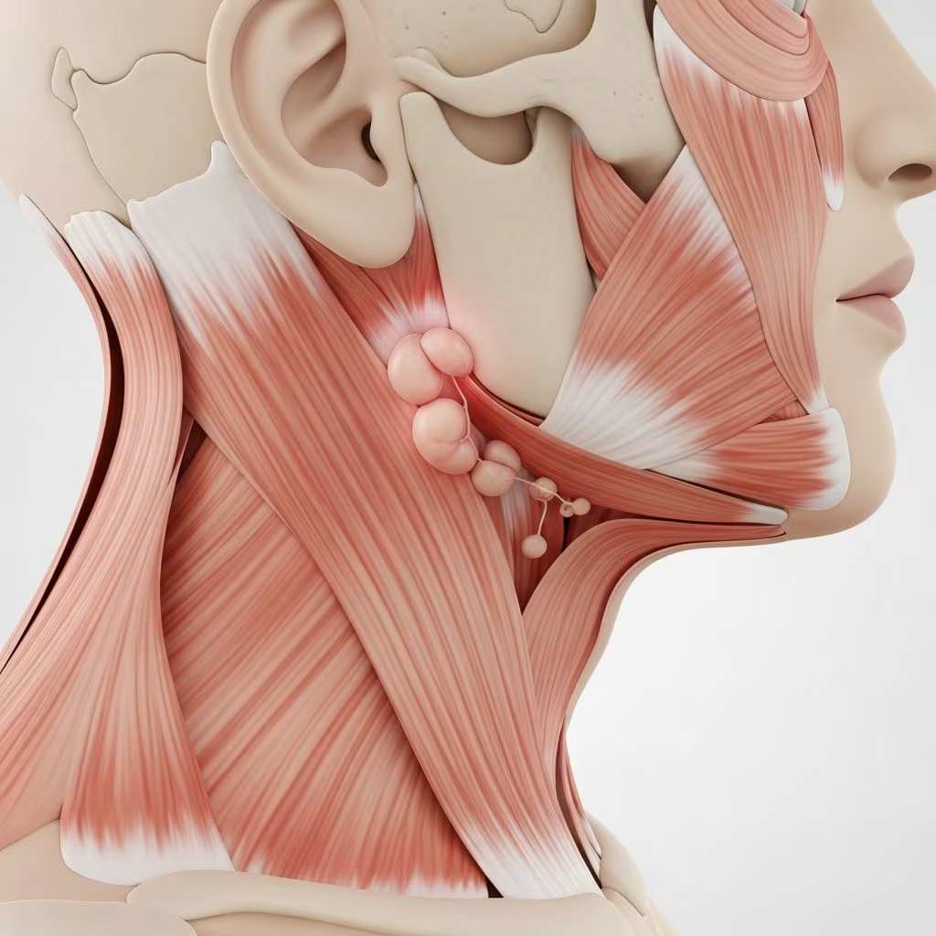
Kimura disease (KD) is a rare, benign, chronic inflammatory disorder primarily affecting the subcutaneous tissues, salivary glands, and lymph nodes, most commonly in the head and neck region. First described in 1937 by Kimm and Szeto as ‘eosinophilic hyperplastic lymphogranuloma’ and later named after Kimura et al. in 1948, KD is characterized by painless subcutaneous nodules or masses, regional lymphadenopathy, marked peripheral eosinophilia, and significantly elevated serum IgE levels. This condition predominantly impacts young Asian males, though cases in other demographics have been reported. The disease mimics neoplastic processes, often leading to diagnostic challenges, but histopathology confirms its benign, reactive nature.
Despite its rarity, KD’s distinct clinicopathological features make it recognizable once suspected. It is not associated with systemic malignancy but can recur and lead to complications like renal involvement. Understanding KD is crucial for dermatologists, pathologists, and ENT specialists to avoid unnecessary aggressive interventions.
Demographics
Kimura disease shows a strong predilection for young Asian males, typically aged 20–40 years, with a male-to-female ratio ranging from 3:1 to 9:1 across studies. It is most prevalent in East Asian populations, particularly Chinese, Japanese, and Korean individuals, though sporadic cases occur in non-Asians, including Caucasians and Africans. Pediatric cases are rare but documented, often presenting similarly to adults.
- Age: Mean onset around 30 years; range 10–70 years.
- Gender: Overwhelmingly male (80–90% of cases).
- Ethnicity: Asians comprise >90% of reported cases.
- Geography: Endemic in Asia; rare elsewhere, suggesting possible genetic or environmental factors.
This demographic skew underscores the need for heightened suspicion in at-risk groups presenting with head/neck masses.
Causes
The exact etiology of Kimura disease remains unknown, but it is hypothesized to involve an aberrant immune response, possibly triggered by allergic, autoimmune, or infectious processes. Key proposed mechanisms include:
- Th2-mediated immunity: Increased Th2 cells and cytokines (IL-4, IL-5, IL-13) drive eosinophilia and IgE production.
- Allergic hypersensitivity: Elevated IgE suggests type I hypersensitivity; many patients have personal/family atopy history.
- Autoimmune factors: Associations with IgG4-related disease (IgG4-RD) in some cases, with overlapping histopathology.
- Infectious triggers: Parasitic infections (e.g., Toxocara) proposed but unproven; no consistent pathogen identified.
- Genetic predisposition: Higher incidence in Asians hints at genetic susceptibility.
Mast cell activation via IgE may contribute to pruritus and inflammation. Trauma or self-antigen exposure could initiate the dysregulated response. No definitive cause is established, classifying KD as idiopathic.
Clinical Features
Patients typically present with painless, firm, ill-defined subcutaneous nodules or masses in the head and neck (75–90% of cases), measuring 1–7 cm in diameter. Common sites include preauricular, postauricular, submandibular, and parotid regions. Regional lymphadenopathy is present in 75–80% of cases, often cervical.
- Skin lesions: Subdermal masses with normal overlying skin or mild hyperpigmentation; pruritus in 10–40%.
- Lymph nodes: Enlarged, rubbery, non-tender.
- Salivary glands: Parotid involvement in 20–40%; painless swelling.
- Other sites: Orbit (proptosis, diplopia), oral cavity, extremities (rare).
- Atypical: Eczema, urticaria, prurigo nodularis; systemic symptoms (fever, weight loss) uncommon (<10%).
Laboratory hallmarks: Peripheral eosinophilia (30–70% WBC), serum IgE >10,000 IU/mL, elevated IgG4 in subsets. Lesions grow slowly or stabilize; spontaneous regression rare.
Complications
Though benign, KD can lead to:
- Nephropathy: Membranous glomerulonephritis or nephrotic syndrome in 10–60% of cases, potentially preceding skin lesions; monitor renal function.
- Recurrence: 20–60% post-treatment.
- Cosmetic/functional: Facial asymmetry, salivary dysfunction, orbital compression.
- Misdiagnosis delay: Mimics lymphoma, leading to overtreatment.
- Rare: IgG4-RD overlap, multiorgan involvement.
Diagnosis
Diagnosis integrates clinical, laboratory, and histopathological findings; biopsy is essential.
Investigations
- Labs: CBC (eosinophilia), serum IgE, IgG4, renal panel, urinalysis.
- Imaging: Ultrasound/CT/MRI for mass extent; avid on PET (mimics malignancy).
- Biopsy: Core/excisional of mass/lymph node.
Histopathology
Characteristic: Follicular hyperplasia, eosinophilic infiltrates/abscesses, postcapillary venule proliferation, fibrosis, Warthin-Finkeldey giant cells, IgE deposits in germinal centers.
| Feature | Description |
|---|---|
| Follicular Hyperplasia | Prominent, reactive germinal centers with eosinophilic folliculolysis. |
| Eosinophils | Diffuse interfollicular infiltrates, microabscesses. |
| Vasculature | Proliferated postcapillary venules with sclerosis. |
| Other | Polkaryocytes, hyalinized collagen, mast cells. |
Immunohistochemistry: CD3+, CD4+, CD20+; polyclonal; distinguishes from lymphoma.
Differential Diagnoses
Key mimics include neoplastic and inflammatory conditions.
| Entity | Clinical | Histopathology | IHC/Key |
|---|---|---|---|
| Kimura Disease | Head/neck masses, LAD, eos, IgE↑ | Follicular hyperplasia, eos abscesses, venules | IgE in GCs |
| Epithelioid Hemangioma | Head/neck papules, pruritus | Vascular proliferation, hobnail ECs, eos | FOSB+ |
| Angiolymphoid Hyperplasia (ALHE) | Superficial erythematous papules, bleed/pruritus | Dermal vessels, lymphoid infiltrate | Female>male |
| Lymphoma (T-FHCL) | Generalized LAD, B-symptoms | Effaced architecture, atypical cells | PD1+, monoclonal |
| Langerhans Cell Histiocytosis | Scalp rash, bone/LAD | CD1a+, S100+ dendritic cells | Birbeck granules |
| Parotid Tumors | Salivary mass | Epithelial proliferation | Cytokeratin+ |
Treatment
No standardized therapy; options based on extent/symptoms.
- Observation: For stable, asymptomatic lesions.
- Surgery: Excision for localized disease; high recurrence (30–60%).
- Corticosteroids: First-line systemic (prednisone 0.5–1 mg/kg); response in 70–80%, but relapse on taper.
- Radiotherapy: For recurrent/unresectable; 20–30 Gy effective.
- Immunosuppressants: Cyclosporine, methotrexate for steroid-resistant.
- Biologics: Mepolizumab (anti-IL5) promising for eosinophilia.
- Other: Topical steroids for skin-limited.
Outcome
Benign with excellent prognosis; no malignant transformation. Recurrence common (25–60%), especially if incomplete excision. Nephropathy may persist/resolve variably; long-term follow-up needed. Spontaneous remission rare; disease often self-limiting over years.
Frequently Asked Questions (FAQs)
Q: Who is most at risk for Kimura disease?
A: Young Asian males aged 20–40 years are most commonly affected, comprising the majority of cases.
Q: Is Kimura disease cancerous?
A: No, it is a benign inflammatory disorder, not neoplastic, confirmed by polyclonal histopathology.
Q: What are the main symptoms?
A: Painless head/neck subcutaneous nodules, lymphadenopathy, pruritus, eosinophilia, and high IgE.
Q: How is it diagnosed?
A: By biopsy showing follicular hyperplasia, eosinophilic infiltrates, and elevated serum IgE/eosinophils.
Q: Does it recur after treatment?
A: Yes, recurrence rates are 20–60%, particularly after surgery or steroid taper.
Q: Can it affect the kidneys?
A: Yes, up to 60% develop nephropathy like membranous glomerulonephritis; screen routinely.
References
- A Comprehensive Review of Kimura Disease — PMC – NIH. 2024. https://pmc.ncbi.nlm.nih.gov/articles/PMC12185846/
- Kimura’s Disease — EyeWiki (AAO). 2023-10-27. https://eyewiki.org/Kimura’s_Disease
- Kimura Disease — DermNet NZ. 2023. https://dermnetnz.org/topics/kimura-disease
- Kimura’s disease: A clinicopathological study of 23 cases — Frontiers in Medicine. 2022-11-11. https://www.frontiersin.org/journals/medicine/articles/10.3389/fmed.2022.1069102/full
- Kimura Disease — Lester D.R. Thompson, MD (AJSP). 2004. https://lesterthompsonmd.com/pdf/AJSP-2004-04_Kimura%20Disease.pdf
- Kimura disease retrospective analysis of 53 cases and three mepolizumab — Dove Press (JIR). 2023. https://www.dovepress.com/kimura-disease-retrospective-analysis-of-53-cases-and-three-mepolizuma-peer-reviewed-fulltext-article-JIR