
Differential diagnosis on histopathology
| Condition | Key Features | Distinguishing from Leishmaniasis |
|---|---|---|
| Leprosy | Perineural granulomas, AFB | No kinetoplast; Fite stain negative for amastigotes |
| Histoplasmosis | Smaller yeasts (2–4 µm), narrow-based budding | GMS positive; kinetoplast absent |
| Post-kala-azar dermal leishmaniasis | Superficial macrophage infiltrates | Amastigotes present; clinical history key |
| Sporotrichosis | Asteroid bodies, yeasts | Cigar-shaped yeasts; culture confirms |
Immunohistochemistry (e.g., anti-Leishmania antibodies) and PCR enhance specificity.
Investigations for leishmaniasis
- Microscopy: Giemsa-stained smears from lesion edges (CL/ML), bone marrow/spleen (VL); sensitivity 50–90%.
- Culture: Novy-MacNeal-Nicolle (NNN) medium; gold standard but slow.
- Molecular: PCR for species ID; highest sensitivity.
- Serology: rK39 for VL; limited for CL.
What is the treatment for leishmaniasis?
Treatment varies by form, species, and region. Consult IDSA/WHO guidelines.
- CL: Local heat/cryotherapy, intralesional antimonials, topical paromomycin; systemic pentavalent antimonials, miltefosine, amphotericin B.
- ML: Liposomal amphotericin B, antimonials.
- VL: Liposomal amphotericin B (first-line), miltefosine.
Related topics on DermNet NZ
- Cutaneous leishmaniasis
- Mucosal leishmaniasis
- Visceral leishmaniasis
- Sandfly bites
Frequently Asked Questions (FAQs)
Q: What does leishmaniasis look like under the microscope?
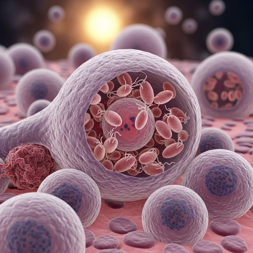
A: Amastigotes are 2–4 µm oval bodies with nucleus and kinetoplast in macrophages; Giemsa stain highlights purple kinetoplasts.
Q: How is leishmaniasis diagnosed pathologically?
A: By identifying amastigotes in tissue smears/biopsies, confirmed by IHC/PCR. Bone marrow for VL.
Q: Which leishmaniasis form has the most parasites histologically?
A: Early CL and VL show heavy parasitisation; late CL/granulomatous forms have fewer.
Q: Can histopathology distinguish Leishmania species?
A: Not reliably; requires PCR or culture for species ID, crucial for ML risk.
Q: Is leishmaniasis contagious person-to-person?
A: No, only via sandfly vectors; blood transfusion rarely.
Word count: 1678 (excluding metadata and references).
References
- ASTMH/IDSA Clinical Practice Guidelines for the Diagnosis and Management of Leishmaniasis — Infectious Diseases Society of America. 2023-05-17. https://www.idsociety.org/practice-guideline/leishmaniasis/
- Leishmaniasis – Infectious Diseases — Merck Manuals Professional Edition. 2024-01-02. https://www.merckmanuals.com/professional/infectious-diseases/extraintestinal-protozoa/leishmaniasis
- Leishmaniasis Fact Sheet — World Health Organization. 2023-03-16. https://www.who.int/news-room/fact-sheets/detail/leishmaniasis
- Leishmaniasis – Symptoms, Diagnosis and Treatment — BMJ Best Practice. 2025-10-15. https://bestpractice.bmj.com/topics/en-us/527
- Leishmaniasis – StatPearls — NCBI Bookshelf, NIH. 2023-07-17. https://www.ncbi.nlm.nih.gov/books/NBK531456/
- Clinical Testing and Diagnosis for Leishmaniasis — Centers for Disease Control and Prevention. 2024-06-11. https://www.cdc.gov/leishmaniasis/hcp/diagnosis/index.html
- Review of the Clinical Presentation, Pathology, Diagnosis, and Treatment of Leishmaniasis — Oxford Academic, Laboratory Medicine. 2023-10-20. https://academic.oup.com/labmed/article/54/4/363/6873137

Medha Deb
Health Writer
This article is for general informational purposes only and is not a substitute for professional medical advice, diagnosis, or treatment. Always seek the advice of a qualified healthcare provider with questions about a medical condition.


