
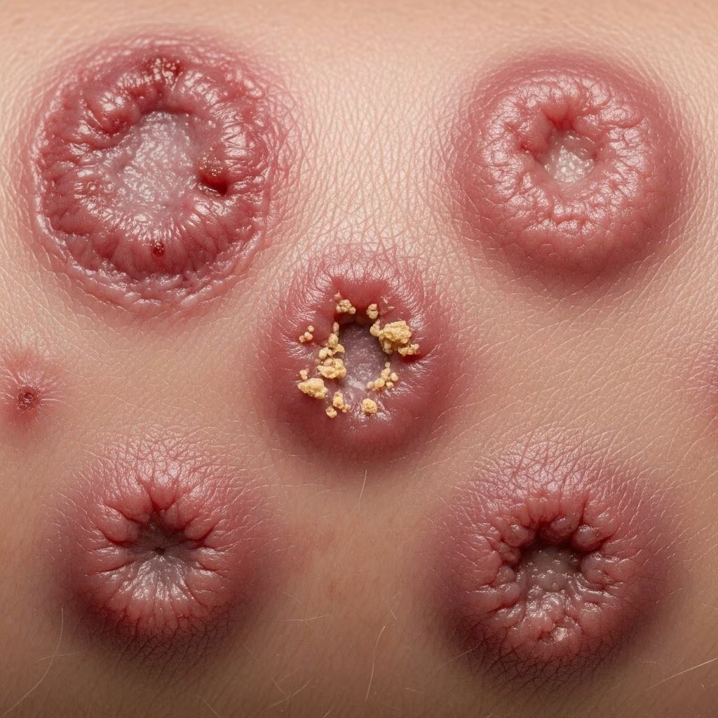
Leishmaniasis is a parasitic disease caused by protozoan parasites of the genus Leishmania, transmitted through the bites of infected female sandflies. It manifests primarily as cutaneous leishmaniasis (CL), the most common form, producing skin ulcers on exposed areas that can scar lifelong. Other forms include mucosal, visceral, and post-kala-azar dermal leishmaniasis (PKDL). This image gallery illustrates diverse clinical presentations across Old World and New World species, aiding in recognition, diagnosis, and management.
What is leishmaniasis?
Leishmaniasis affects over 90 countries, with 95% of cutaneous cases in the Americas, Mediterranean, Middle East, and central Asia. The WHO Eastern Mediterranean region reports 80% of global CL cases. Parasites infect macrophages, leading to localized or systemic disease. Cutaneous forms begin as papules at bite sites, evolving into ulcers with raised violaceous borders over 2-4 weeks incubation. Visceral leishmaniasis (VL), caused by L. donovani and L. infantum, involves fever, splenomegaly, and high mortality if untreated.
Skin lesions vary by species: Old World (L. major, L. tropica) often self-heal in 2-5 years, while New World (L. braziliensis) risks mucosal spread. Diffuse cutaneous leishmaniasis (DCL) features widespread hypopigmented plaques, resistant to immunity.
Leishmaniasis images – codes and images explained
This collection features annotated images of lesions from various Leishmania species. Codes indicate geography (Old World: OW; New World: NW), species, and lesion type. Images depict progression from early papules to chronic ulcers, satellite lesions, and complications like secondary infection or scarring. Visual identification is crucial as symptoms mimic other dermatoses.
Cutaneous leishmaniasis images
Cutaneous leishmaniasis (CL) dominates, with lesions on exposed skin like legs, arms, and face. Initial painless nodules ulcerate centrally, forming volcano-like craters with indurated borders and crusts. Healing leaves atrophic scars. Multiple lesions occur from simultaneous bites.
- OW L. major: Rapidly ulcerating papules on legs, healing with depigmented scars. Common in Middle East, Afghanistan.
- OW L. tropica: Chronic nodules on face/arms, urban anthroponotic form in Syria, Saudi Arabia.
- NW L. mexicana: Chiclero ulcer on ears, slow-healing in Mexico/Guatemala forest workers.
- NW L. braziliensis: Multiple ulcers risking espundia (mucosal extension).
Satellite lesions and lymphadenopathy (chiclero ear) are hallmarks. In immunosuppressed patients, lesions disseminate.
Diffuse cutaneous leishmaniasis images
Diffuse cutaneous leishmaniasis (DCL) arises in anergic hosts, infecting L. aethiopica (OW, Ethiopia) or L. mexicana (NW). Non-ulcerative, symmetric papules/nodules evolve into plaques across face, ears, trunk, sparing mucosa initially. Leonine facies from ear involvement mimics leprosy. Lesions persist years, hypopigmented, with myriad amastigotes.
Images show butterfly distribution on cheeks, nodular ears, extensor limb plaques. No spontaneous healing; requires systemic therapy.
Mucocutaneous leishmaniasis images
Mucocutaneous leishmaniasis (MCL or espundia) follows NW CL, especially L. braziliensis, via lymphatic/hematogenous spread. Onset months to decades post-skin healing, destroying nasal, oral, pharyngeal mucosa. Images reveal nasal septum perforation, palatal ulcers, lip/nose erosion causing disfigurement, stigma.
Severe cases erode soft/hard palate, leading to aspiration, death if untreated. Early reddish granulations progress to vegetating masses.
Post-kala-azar dermal leishmaniasis (PKDL) images
PKDL complicates VL treatment/recovery, prevalent in India/Sudan (5-10% cases). Maculopapular rash on face, arms, trunk months-years post-VL. Hypopigmented macules, nodules; severe Sudanese form invades mucosa.
Images depict perioral papules, trunk patches, serving as VL reservoir. Self-limited in Indians, chronic in Africans.
Visceral leishmaniasis with skin lesions
VL primarily systemic but post-therapy skin rashes occur as PKDL. Rare primary cutaneous signs include facial papules. Images show hyperpigmented macules, sparse papules in VL patients.
Leishmaniasis on the lip images
Facial CL/MCL frequently affects lips: eroded vermilion, crusted ulcers from L. tropica or L. braziliensis. Progression to cheilitis mutilans with scarring.
Treatment images
Treatment visuals: Pre/post intralesional antimonials (sodium stibogluconate) shrinking ulcers; cryotherapy blistering edges; thermotherapy red induration. Systemic miltefosine, amphotericin B for complex cases. Scars persist despite healing.
Frequently Asked Questions
What does leishmaniasis look like?
Skin-colored papule enlarges to ulcer with raised violaceous border, central crust/depression. Volcano-shaped, painless unless superinfected.
How do you get leishmaniasis?
Bite from Phlebotomus (OW) or Lutzomyia (NW) sandflies, 2-3mm, dusk-biting. Reservoirs: rodents, dogs, humans.
Where is leishmaniasis found?
Tropics/subtropics: 90+ countries. CL hotspots: Syria, Afghanistan, Brazil; VL: India, East Africa.
Does leishmaniasis go away on its own?
Localized CL often self-resolves in months-years, scarring. DCL, MCL, VL do not; treatment essential.
How is leishmaniasis diagnosed from images?
Clinical: ulcer morphology, travel history. Confirm via biopsy smear (amastigotes), PCR. RDT available but limited.
Table: Leishmania Species and Lesion Characteristics
| Species | Region | Lesion Type | Key Features |
|---|---|---|---|
| L. major | OW | Localized CL | Multiple moist ulcers, self-healing |
| L. tropica | OW | Localized CL | Chronic dry ulcers, recidivans |
| L. aethiopica | OW | DCL | Diffuse nodules, leonine face |
| L. mexicana | NW | CL/DCL | Chiclero ear, hypopigmented |
| L. braziliensis | NW | CL/MCL | Ulcers + mucosal destruction |
| L. donovani/infantum | Global | VL/PKDL | Systemic + skin rash |
This table summarizes species-specific presentations, guiding differential diagnosis.
Leishmaniasis images underscore diagnostic challenges: mimic pyoderma, sporotrichosis, basal cell carcinoma. Travel/endemic exposure history pivotal. Prevention: Insecticide nets, repellents. Vaccine trials ongoing. Complex cases need specialist referral; disfigurement impacts psychosocial health.
References
- Leishmaniasis – StatPearls — Arango Dukue C, et al. National Center for Biotechnology Information (NCBI). 2023-07-17. https://www.ncbi.nlm.nih.gov/books/NBK531456/
- Leishmaniasis: Causes, Symptoms, Diagnosis & Treatment — Cleveland Clinic. 2023-08-01. https://my.clevelandclinic.org/health/diseases/24539-leishmaniasis
- Leishmaniasis Fact Sheet — World Health Organization (WHO). 2023-03-02. https://www.who.int/news-room/fact-sheets/detail/leishmaniasis
- Cutaneous Leishmaniasis Facts — Drugs for Neglected Diseases initiative (DNDi). 2024-01-15. https://www.dndi.org/diseases/cutaneous-leishmaniasis/facts/
- Cutaneous Leishmaniasis: Recognition and Treatment — Herwaldt BL. American Academy of Family Physicians (AAFP). 2004-03-15. https://www.aafp.org/pubs/afp/issues/2004/0315/p1455.html
- Leishmaniasis — Pan American Health Organization (PAHO/WHO). 2023-11-20. https://www.paho.org/en/topics/leishmaniasis



