
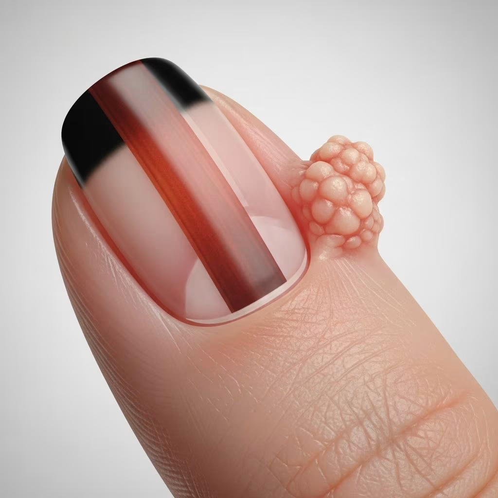
Onychopapilloma is a rare benign neoplasm primarily affecting the nail matrix and nail bed. It typically presents as an isolated lesion on a single nail, most commonly the thumb, manifesting as longitudinal chromonychia—often erythronychia (red streak)—extending from the lunula to the distal nail edge, accompanied by subungual hyperkeratosis and frequent onycholysis.
Introduction
Onychopapilloma represents the most common cause of monodactylous longitudinal erythronychia, a narrow linear streak of red discoloration under the nail plate originating from the proximal nail matrix. First described in 1995 by Baran and Perrin, this entity is characterised by papillomatous proliferation of the nail bed epithelium with matrix metaplasia. While overwhelmingly benign, rare malignant transformation (e.g., Bowen’s disease histology) has been reported, and multiple lesions raise concern for BAP1 tumour predisposition syndrome.
The lesion arises from the distal nail matrix and subungual tissues, producing a keratotic papillary growth that disrupts normal nail architecture. Clinically, it interferes with fine motor tasks due to mechanical friction and may cause cosmetic distress. Early recognition prevents misdiagnosis as malignancy, such as subungual melanoma or squamous cell carcinoma.
Demographics
Onychopapilloma predominantly affects middle-aged adults, with a noted predilection for females. It most frequently involves a single digit, particularly the thumb (most common), followed by the index finger. Multiple nail involvement is exceptional (<1% of cases) and strongly associated with BAP1-TPDS, observed in 88% of affected individuals over age 30.
- Age: Typically 40–60 years; rare in children.
- Sex: Female predominance (ratio ~2:1).
- Site: Thumb > index finger > other fingernails; toenails less common.
- Distribution: Unidigital (95%); polydigital rare, syndromic.
Causes
The aetiology of onychopapilloma remains unknown. Proposed mechanisms include:
- Neoplastic hyperplasia: Papillomatous acanthosis of nail bed epithelium, possibly triggered by chronic microtrauma or irritation.
- Reactive process: Secondary to inflammatory nail disorders (e.g., lichen planus).
- Genetic predisposition: Germline BAP1 mutations in cases of multiple onychopapillomas, linking to hereditary cancer syndrome with mesothelioma, uveal/renal cell carcinoma risks.
- Excluded associations: No definitive link to HPV, fungi, or other infections despite historical speculation.
Histologically, uniform eosinophilic epithelial cells with hyperkeratosis confirm benign papilloma; matrix involvement causes the characteristic longitudinal streak.
Clinical features
The classic presentation combines longitudinal chromonychia with distal subungual hyperkeratosis (keratotic papule), visible as a white/yellow mass under the free edge causing V-shaped notching or onycholysis.
| Feature | Description | Frequency |
|---|---|---|
| Chromonychia | Narrow (0.3–1.5 mm) streak: red (erythronychia), white (leukonychia), or brown (melanonychia) | ~100% |
| Hyperkeratosis | Distal subungual papule; layered keratin ± hemorrhage | 90–95% |
| Onycholysis/splitting | Distal detachment; V-notch fissure | 70–80% |
| Splinter haemorrhages | Linear red-brown streaks along band | 50–60% |
| Pain | Dull ache on pressure/picking objects | 20–40% |
Dermoscopy reveals a linear red streak with distal keratotic plug, splinter-like haemorrhages, and V-shaped notch—highly suggestive. Progression is slow; width may increase over years.
Diagnosis
Diagnosis relies on correlating clinical, dermoscopic, and histopathologic findings. Suspect onychopapilloma in solitary longitudinal erythronychia with hyponychial hyperkeratosis.
- Clinical exam: Inspect for keratotic papule, onycholysis, haemorrhages.
- Dermoscopy: Confirms linear morphology, distal keratoma.
- Nail clipping/biopsy: Essential if enlarging, painful, or atypical. Histology shows papillomatous acanthosis, eosinophilic matrix metaplasia, focal hyperkeratosis ± haemorrhage.
- Imaging: Rarely, ultrasound/MRI for deep extension.
Multiple lesions mandate BAP1 genetic testing.
Differential diagnoses
Onychopapilloma mimics serious pathology, necessitating biopsy for longitudinal chromonychia >1 mm or enlarging.
| Differential | Key Distinguishers |
|---|---|
| Subungual melanoma | Irregular pigmentation, Hutchinson sign, rapid growth, periungual pigmentation |
| Squamous cell carcinoma | Polydactylous, periungual invasion, pain/bleeding, destructive |
| Wart (HPV) | Multiple specks, periungual verrucae, contagious |
| Gliomatrixoma/Onychomatricoma | Thick spongiotic nail, ‘doughnut’ dermoscopy, no hyperkeratosis |
| Lichen planus/Darier | Polydactylous, mucosal/skin involvement precedes |
| BAP1-TPDS | Multiple nails, family cancer history |
Treatment
Asymptomatic lesions require observation with photography. Intervention for pain, progression, or diagnostic uncertainty:
- Conservative: Nail avulsion + curettage (diagnostic/therapeutic).
- Surgical: Complete excision (matrix + bed) prevents recurrence; Mohs micrographic surgery for margins.
- Laser/ablative: CO2 laser vaporisation reported successful in select cases.
- BAP1 cases: Genetic counselling, cancer surveillance.
Recurrence is low post-excision (~5%).
Outcome
Excellent prognosis: benign, non-metastasising. Surgical removal resolves symptoms and confirms diagnosis. Rare malignant variants require oncologic management. In BAP1-TPDS, onychopapilloma signals high lifetime cancer risk (mesothelioma >40%, melanomas >15%); early screening vital.
Patient education reduces anxiety over ‘nail cancer’ fears. Regular follow-up (6–12 months) for monodactylous cases; annual for multiples.
Frequently Asked Questions
What is onychopapilloma?
A rare benign tumour of the nail matrix/bed causing longitudinal red/white streaks and distal thickening.
Is onychopapilloma cancerous?
Typically no; exceedingly rare malignant change reported. Biopsy if suspicious.
Does it hurt?
Often asymptomatic; some experience dull pain on pressure or fine tasks.
Why my thumbnail?
Thumb most common site due to trauma exposure; usually solitary.
Can it spread to other nails?
Solitary typical; multiple suggests BAP1 syndrome—needs genetic eval.
Treatment necessary?
Observe if asymptomatic; excise for pain/doubt.
Will it grow back?
Rare post-excision; complete removal curative.
References
- Onychopapilloma: Symptoms & Management — Medicover Hospitals. 2024. https://www.medicoverhospitals.in/diseases/onychopapilloma/
- A Rare Case of Onychopapilloma Presenting as a Longitudinal Erythronychia — PMC/NCBI (peer-reviewed). 2021-03-15. https://pmc.ncbi.nlm.nih.gov/articles/PMC7992634/
- Onychopapilloma Patient Information Leaflet — British Hair and Nail Society. 2021. https://bhns.org.uk/uploads/pages_media/605786839_0_OnychopapillomaPIL.pdf
- Onychopapilloma: A Potential Mimicker — Actas Dermo-Sifiliográficas (peer-reviewed). 2024. https://www.actasdermo.org/es-onychopapilloma-a-potential-mimicker-articulo-S0001731024000504
- Onychopapilloma — DermNet NZ (official dermatology resource). 2024. https://dermnetnz.org/topics/onychopapilloma
- Benign nail condition linked to rare syndrome — National Institutes of Health (NIH.gov). 2022-11-28. https://www.nih.gov/news-events/news-releases/benign-nail-condition-linked-rare-syndrome-greatly-increases-cancer-risk
- Onychopapilloma: its clinical, dermoscopic and pathologic features — Journal of the European Academy of Dermatology and Venereology (Wiley, peer-reviewed). 2023. https://onlinelibrary.wiley.com/doi/10.1111/jdv.18461



