
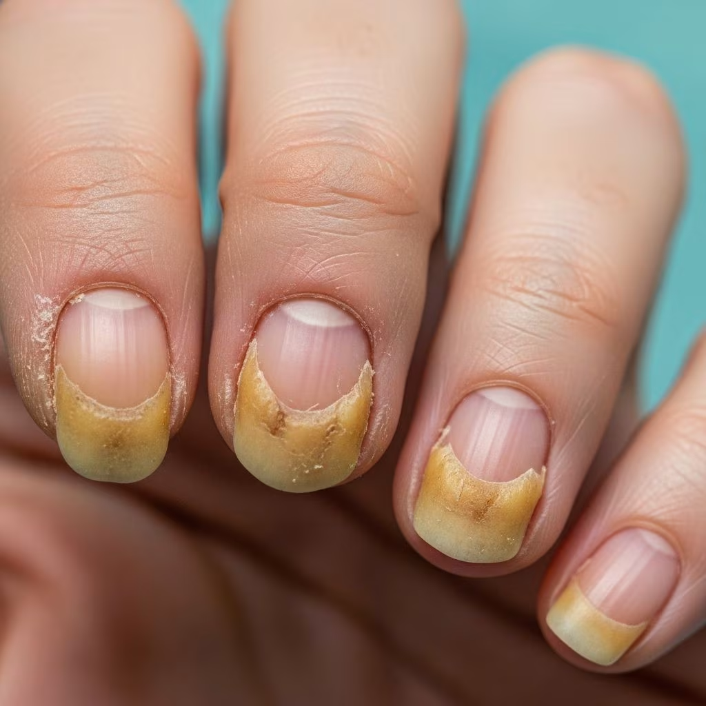
Pachyonychia congenita (PC) is a group of rare, inherited disorders of keratinisation characterised by thickened skin on the palms and soles (palmoplantar keratoderma), hypertrophic nail dystrophy, and white patches in mucous membranes (leukokeratosis). The condition profoundly impacts quality of life due to severe pain, particularly on weight-bearing areas of the feet.
Introduction
Pachyonychia congenita involves defects in the process by which keratin—a tough protein—is formed and deposited in the skin’s outermost layer (epidermis). Mutations in specific keratin genes disrupt the structural integrity of skin cells, leading to fragility under mechanical stress. Normal activities like walking cause cell breakdown, resulting in blisters, calluses, and nail abnormalities. PC is classified into subtypes based on the affected gene, influencing the predominant clinical features.
Demographics
PC affects approximately 1 in 150,000 to 1 in 1,000,000 individuals worldwide, with no strong racial or geographic predilection. It manifests at birth or early childhood, often noticed when infants begin weight-bearing. Females and males are equally affected due to autosomal dominant inheritance. Natal teeth occur more frequently in PC-17 subtype.
- Prevalence: Rare; exact figures vary but consistently reported as under 1 per 100,000.
- Age of onset: Nails dystrophic by 1 year; plantar keratoderma with walking (1-2 years).
- Sex distribution: Equal.
Causes
PC results from autosomal dominant mutations in five keratin genes: KRT6A (PC-6a), KRT6B (PC-6b), KRT6C (PC-6c), KRT16 (PC-16), and KRT17 (PC-17). These encode high-sulfur keratins expressed in epithelial tissues like nails, palms, soles, oral mucosa, hair follicles, and glands. Mutant keratins form unstable filaments, rendering cells susceptible to trauma-induced rupture.
| Subtype | Gene | Key Features |
|---|---|---|
| PC-6a | KRT6A | Focal plantar keratoderma, oral leukokeratosis, cysts |
| PC-6b | KRT6B | Palmoplantar keratoderma, oral involvement |
| PC-6c | KRT6C | Mild skin/nail changes, focal hyperkeratosis |
| PC-16 | KRT16 | Palmoplantar pain, nail dystrophy, follicular hyperkeratosis |
| PC-17 | KRT17 | Natal teeth, oral leukokeratosis, steatocystomas |
De novo mutations account for ~30% of cases; penetrance is high (>95%), but expressivity varies.
Clinical Features
The hallmark triad is hypertrophic nail dystrophy, painful palmoplantar keratoderma, and mucosal leukokeratosis. Plantar pain is debilitating, often requiring mobility aids.
Nail Dystrophy
Toenails show wedge-shaped thickening, subungual hyperkeratosis, and yellow-brown discoloration, starting infancy. Fingernails less severely affected. Upward nail plate angle >10° is characteristic.
Palmoplantar Keratoderma
Firm, painful calluses on pressure points (heels, balls of feet) form with ambulation. Blisters beneath cause excruciating pain; hyperhidrosis worsens friction. Hands show milder hyperkeratosis.
Oral Leukokeratosis
White plaques on tongue, gingivae, buccal mucosa from birth/infancy. Benign, thickens with age; PC-6a most severe.
Cysts
Steatocystomas (PC-17), vellus hair cysts (PC-6a), hidrocystomas, odontogenic cysts. Appear post-puberty.
Other Features
- Follicular hyperkeratosis: On legs, arms (PC-16).
- Hyperhidrosis: Palms/soles (~50%).
- Natal/prenatal teeth: PC-17 (~50%).
- Rare: Angular cheilitis, hoarse voice, first bite syndrome (jaw pain on eating).
Classification
Originally Jadassohn-Lewandowsky (PC-1: nails/skin/mucosa) vs. Jackson-Lawler (PC-2: cysts/natal teeth). Now gene-based subtypes (PC-6a/b/c, PC-16/17) better reflect phenotypes.
Diagnosis
Suspected clinically: nail thickening + plantar keratoderma/pain by age 10 in 97%. Confirmed by genetic testing identifying pathogenic variant in KRT6A/B/C/16/17. Skin biopsy shows compact hyperkeratosis, acanthosis; electron microscopy reveals keratin aggregates (research use).
- 97% sensitivity: Triad by childhood.
Differential Diagnoses
| Condition | Distinguishing Features |
|---|---|
| Unna-Thost PPK | Diffuse non-painful keratoderma; no nail/mucosal changes |
| Vörner PPK | Painful but no cysts/nails |
| Onychogryphosis | Acquired claw-like nails; no skin changes |
| Candida onychomycosis | Paronychia without PPK; KOH positive |
| Steatocystoma multiplex | Cysts without nails/PPK |
Rule out infection if isolated nail changes.
Treatment
No cure; symptomatic management focuses on pain relief, preventing complications. Multidisciplinary: dermatology, podiatry, pain specialists.
Mechanical Debridement
Regular podiatrist-performed paring/grinding of calluses/nails essential. Avoid over-thinning to prevent blistering.
Topical Therapies
- Keratolytics: Urea (20-40%), salicylic acid (6-40%) soften hyperkeratosis; limited efficacy.
- Moisturizers: Emollients (petrolatum) reduce fissuring.
- Retinoids: Topical tretinoin thins calluses but irritates.
Systemic Therapies
- Oral retinoids (acitretin): Reduce thickness but increase pain/skin fragility; short courses.
Pain Management
- NSAIDs, opioids pre-activity.
- Botulinum toxin injections: Reduce sweating/pain (lasts 3-6 months).
Devices
Custom orthotics, padded insoles redistribute pressure. Severe cases: crutches/wheelchair.
Infection Control
Antifungals/bacterials for secondary issues.
Experimental
siRNA (targeting mutant keratin), topical rapamycin, mTOR inhibitors in trials. Botulinum, fluorouracil tested.
Outcome
Chronic, lifelong; plantar pain limits mobility/employment. Complications: secondary infection, disability. Early intervention improves function. Genetic counseling crucial (50% recurrence risk). Patient registries (pachyonychia.org) aid research.
Frequently Asked Questions
What is pachyonychia congenita?
A rare genetic disorder causing painful thick calluses on feet, deformed nails, and mouth plaques due to keratin gene mutations.
Is there a cure for PC?
No cure exists; treatments manage symptoms like pain and thickening.
How is PC inherited?
Autosomal dominant: one mutated gene copy from a parent suffices; 30% sporadic.
Does PC affect life expectancy?
No; it impacts quality of life via pain/mobility but not survival.
Can children with PC walk normally?
Often not; pain may require aids from toddlerhood.
References
- Pachyonychia Congenita – Symptoms, Causes, Treatment — NORD (rarediseases.org). 2023. https://rarediseases.org/rare-diseases/pachyonychia-congenita/
- Pachyonychia Congenita – Thick Nails & Calluses — NIAMS/NIH (.gov). 2024-10-15. https://www.niams.nih.gov/health-topics/pachyonychia-congenita
- Pachyonychia Congenita: Diagnosis, Treatment, and Steps to Take — NIAMS/NIH (.gov). 2024. https://www.niams.nih.gov/health-topics/pachyonychia-congenita/diagnosis-treatment-and-steps-to-take
- Discover & Support Pachyonychia Congenita — Rare Genomics Institute. 2023. https://www.raregenomics.org/pachyonychia-congenita
- Pachyonychia Congenita: Clinical Features and Future Treatments — Keio Journal of Medicine (PDF via pachyonychia.org). 2023-09-28. https://www.pachyonychia.org/wp-content/uploads/2024/02/PCClinicalFeaturesKeioJournaladvpub_2023-0012-IR.pdf
- Pachyonychia congenita — MedlinePlus Genetics/NLM (.gov). 2024. https://medlineplus.gov/genetics/condition/pachyonychia-congenita/



