
Panfolliculoma is a rare benign cutaneous adnexal tumour characterized by differentiation towards all components of the hair follicle, from infundibulum to hair bulb. First described in 1991, it represents a unique neoplasm that recapitulates the entire follicular architecture within a well-circumscribed dermal lesion.
Introduction
Panfolliculoma belongs to the spectrum of hair follicle tumours, which are part of the broader category of skin adnexal neoplasms. These tumours arise from the pilosebaceous unit, comprising the hair follicle and associated sebaceous glands responsible for hair production and skin lubrication. Unlike more common follicular tumours such as trichilemmoma or pilomatricoma, panfolliculoma uniquely demonstrates the full spectrum of follicular differentiation, making its pathological recognition critical for accurate diagnosis.
This article details the clinical features, histopathological findings, immunohistochemical profile, differential diagnoses, and management strategies for panfolliculoma. Understanding this entity is essential, as it can mimic more aggressive lesions and requires distinction from malignant counterparts.
Clinical Features
- Incidence: Extremely rare, with only sporadic case reports and small series documented in the literature.
- Age: Predominantly affects adults, typically in the 4th to 7th decades of life.
- Gender: No significant gender predilection; affects men and women equally.
- Site: Most commonly arises on the face (cheek, nose, eyelid) or scalp, though lesions have been reported on the trunk, extremities, and anogenital region.
- Presentation: Appears as a slowly enlarging, asymptomatic papule, nodule, or plaque, ranging from 0.3 to 1.5 cm in diameter. The surface may be smooth, verrucous, or eroded.
- Associations: No known syndromic associations, unlike some other adnexal tumours linked to genetic syndromes such as Birt-Hogg-Dubé or Cowden disease.
Clinically, panfolliculoma is often mistaken for basal cell carcinoma, sebaceous hyperplasia, or other adnexal tumours due to its unremarkable appearance. Complete surgical excision is curative, with no reported recurrences.
Histopathology
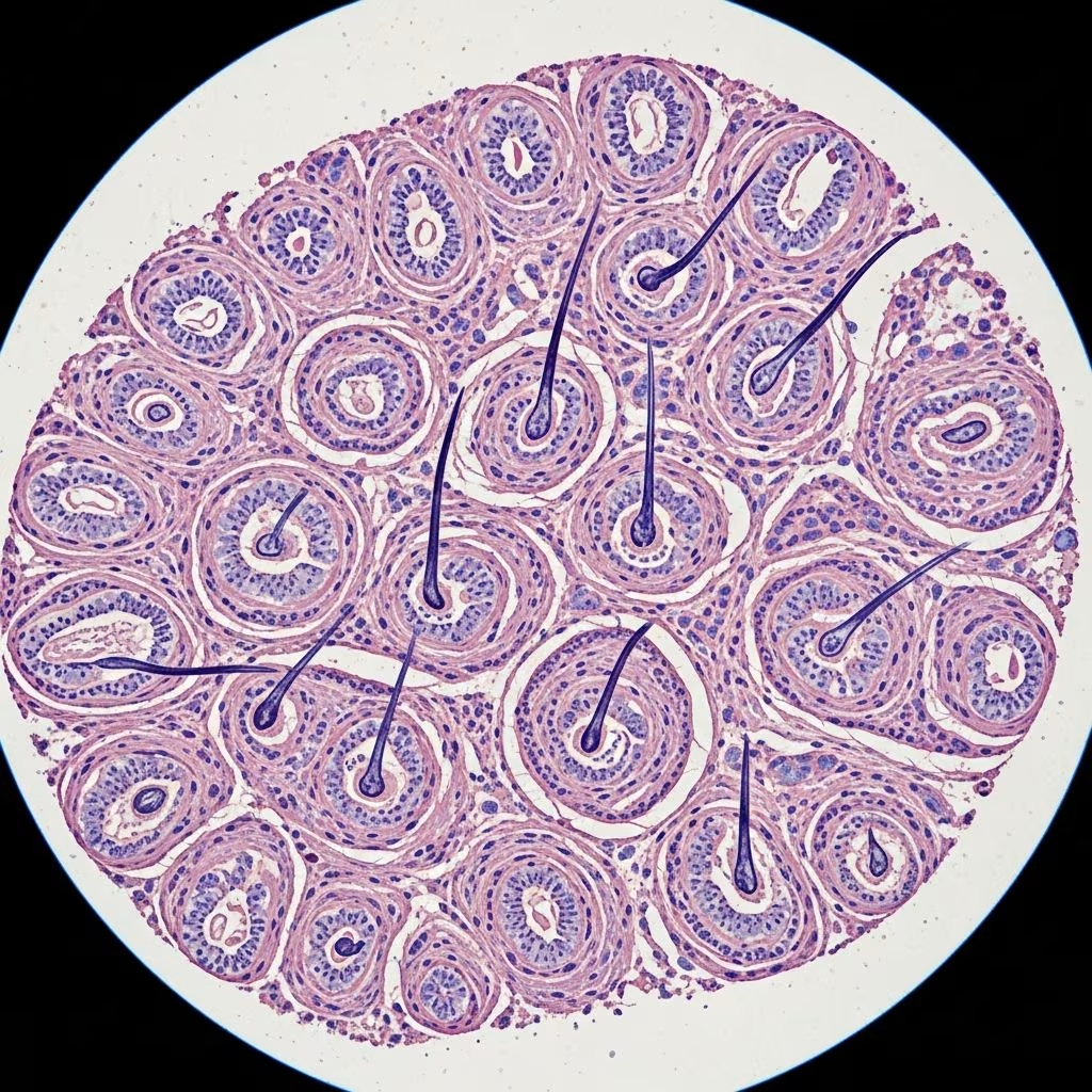
Microscopically, panfolliculoma presents as a well-circumscribed, dermal-based proliferation of epithelial islands and cysts exhibiting multifaceted follicular differentiation. The lesion lacks connection to the epidermis in most cases and is surrounded by a thin fibrous stroma.
Microscopic Description
- Architecture: Closely packed solid nests, cords, and cystic spaces within the dermis, often extending into the subcutis. The tumour is multilobulated and sharply demarcated.
- Infundibular differentiation: Cystic structures lined by stratified squamous epithelium with a prominent granular layer and basket-weave orthokeratotic keratin, mimicking the follicular infundibulum.
- Isthmic differentiation: Areas resembling the inner and outer root sheaths, with pale eosinophilic cuticle and trichohyalin granules.
- Matrical differentiation: Basaloid islands with peripheral palisading, shadow cells (ghost cells), and filamentous keratin production indicative of hair matrix.
- Bulbar differentiation: Larger lobules with papillary mesenchymal bodies (trichogenic stroma) simulating the hair bulb, often with associated hair shaft fragments.
- Additional features: Scattered sebocytes, ductal structures, or Merkel cells may be present, reflecting pilosebaceous or neurofollicular elements. No significant cytological atypia, mitoses, or necrosis.
Variants include pleomorphic (with atypical but benign features), sebaceous, or regressing forms, but all maintain benign behaviour.
Images in Pathology
Low-power view reveals a well-circumscribed dermal nodule composed of variably sized epithelial units (scanning magnification). Higher power demonstrates infundibular cysts with granular layer (Figure 1, akin to DermNet image), matrical lobules with shadow cells, and bulb-like structures with papillary mesenchyme.
Histopathology Images
| Feature | Description | Typical Appearance |
|---|---|---|
| Infundibular cyst | Stratified squamous epithelium with granular layer | Basket-weave keratin |
| Matrical lobule | Basaloid cells transitioning to shadow cells | Peripheral palisading |
| Hair bulb | Papillary mesenchymal body | Matrical epithelium surrounding stroma |
| Root sheath | Trichohyalin granules | Pale cuticle |
Immunohistochemistry
Immunohistochemical studies confirm follicular differentiation and aid in distinguishing panfolliculoma from mimics.
- Cytokeratins: AE1/AE3 positive throughout; high-molecular-weight CKs (CK5/6) in outer root sheath areas; CK15 in bulb regions.
- Trichohyalin: Positive in isthmic/root sheath components.
- β-catenin: Membranous in most areas; nuclear in matrical zones (similar to pilomatricoma but less intense).
- PHLDA1: Positive in shadow cells, supporting matrical differentiation.
- Other markers: Bcl-2 highlights basaloid cells; Ki67 proliferation index low (<5%); negative for BerEP4, EMA (against BCC).
These markers underscore the panfollicular nature without evidence of malignancy.
Differential Diagnosis
Panfolliculoma’s diverse differentiation leads to overlap with other follicular neoplasms. Key discriminators include the presence of all follicular levels.
| Tumour | Key Features | Distinguishing from Panfolliculoma |
|---|---|---|
| Trichoblastoma | Basaloid nests with fibroblastic stroma, fibroepithelial units | Lacks infundibulum, bulb, advanced sheath differentiation |
| Trichoepithelioma | Symmetric, horn cysts, papillary mesenchymal bodies | Predominantly primitive follicular; no full bulb/matrical |
| Pilomatricoma | Predominantly shadow cells, basophilic matrix | Limited to matrical differentiation only |
| Follicular Poroma/Pilomatrical Carcinoma | Connected to epidermis, atypia, infiltration | Malignant features absent in panfolliculoma |
| Basal Cell Carcinoma | Retraction artifact, BerEP4+, infiltrative | No full follicular recapitulation; atypia present |
Other considerations: trichofolliculoma (central dilated follicle with vellus hairs), fibrofolliculoma (thin epithelial cords in fibrous sheath).
Pathogenesis
The exact aetiology remains unclear, but panfolliculoma likely arises from pluripotent follicular stem cells capable of multidirectional differentiation. Genetic studies are limited, with no recurrent mutations identified, unlike syndromic tumours.
Treatment
- Primary: Complete surgical excision with clear margins, which is curative.
- Follow-up: No routine surveillance needed due to benign nature; monitor for rare local recurrence if incompletely excised.
- Biopsy considerations: Avoid superficial biopsies; ensure adequate depth for full architectural assessment.
Frequently Asked Questions
Q: Is panfolliculoma malignant?
A: No, panfolliculoma is entirely benign with no metastatic potential. Rare atypical variants remain non-aggressive.
Q: How is panfolliculoma diagnosed?
A: Diagnosis relies on histopathology demonstrating all hair follicle components; immunohistochemistry supports but is not essential.
Q: What is the most common location for panfolliculoma?
A: Face and scalp are most frequent sites.
Q: Does panfolliculoma require special follow-up?
A: No, simple excision suffices; no syndromic associations.
Q: Can panfolliculoma recur?
A: Recurrence is exceptional with complete removal.
Conclusion
Panfolliculoma exemplifies the complexity of adnexal pathology, requiring careful microscopic evaluation to appreciate its panfollicular differentiation. Awareness prevents misdiagnosis as malignancy and ensures appropriate conservative management.
References
- Panfolliculoma — Steven D. Billings, MD. Basicmedicalkey.com. Accessed 2026. https://basicmedicalkey.com/panfolliculoma/
- Panfolliculoma and histopathologic variants: a study of 19 cases — PubMed. 2014-04-01. https://pubmed.ncbi.nlm.nih.gov/24698942/
- Hair follicle tumours — DermNet NZ. 2013. https://dermnetnz.org/topics/hair-follicle-tumours
- Trichofolliculoma pathology — DermNet NZ. 2014. https://dermnetnz.org/topics/trichofolliculoma-pathology



