
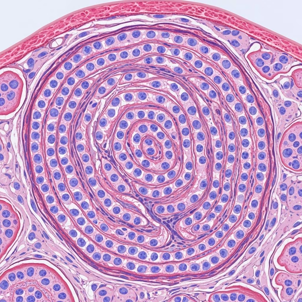
Perineurioma is an uncommon benign peripheral nerve sheath tumour composed exclusively of perineural cells.
Histology of perineurioma
Perineurioma tumour cells are spindle shaped, with long delicate cell processes amidst a collagenous stroma. The spindle cells have pale open nuclei with a delicate chromatin pattern, an inconspicuous eosinophilic nucleolus and indistinct cell borders.
Myxoid change may be seen and can cause diagnostic confusion. Whorls of cells are often seen which are reminiscent of meningioma.
- Key histological features:
- Spindle-shaped tumour cells with elongated, delicate cytoplasmic processes
- Pale, open nuclei with fine chromatin and small nucleoli
- Indistinct cell borders blending into collagenous stroma
- Frequent whorling patterns and storiform architecture
- Variable myxoid or collagenous stromal background
- Absence of axons or residual nerve structures (extraneural type)
Scanning power reveals a well-circumscribed but unencapsulated dermal nodule composed of bland spindle cells arranged in fascicles, whorls, and storiform patterns within a collagenous stroma. Higher magnification demonstrates the characteristic bipolar cytoplasmic processes extending between cells, creating a lace-like pattern. The nuclei show delicate longitudinal nuclear grooves and are notably bland without significant atypia or mitoses.
Subtypes of perineurioma
Perineuriomas are classified into several histological subtypes based on their location and growth patterns:
Intraneural perineurioma
Occurs within nerve fascicles, typically affecting major peripheral nerves in children and young adults. Characterized by concentric “onion bulb” arrangements of perineurial cells around central axons and Schwann cells. Often associated with motor deficits.
Extraneural (soft tissue) perineurioma
Most common type presenting in dermis/subcutis. Lacks association with nerve fascicles and shows complete replacement of normal neural elements. Typical storiform-fascicular growth with collagenous stroma.
Sclerosing perineurioma
Predominantly acral distribution (hands/feet). Features densely collagenized stroma with epithelioid/plump spindle cells arranged in cords and small whorls. Characteristic “platelet” forms with cytoplasmic inclusions.
Reticular perineurioma
Characterized by prominent reticular growth pattern with elongated, wavy spindle cells forming pseudovascular spaces. Often shows myxoid stroma and can mimic low-grade angiosarcoma.
Pseudolipoblastic perineurioma
Rare variant with vacuolated cells resembling lipoblasts. Essential to recognize EMA/claudin-1 positivity to avoid misdiagnosis as liposarcoma. Typically dermal nodules in acral sites.
| Subtype | Location | Key Features | Age Group |
|---|---|---|---|
| Intraneural | Major peripheral nerves | Onion bulb whorls around axons | Children/young adults |
| Extraneural | Dermis/subcutis/soft tissue | Storiform, collagenous stroma | Adults |
| Sclerosing | Acral (hands/feet) | Plump cells, dense collagen | Young adults |
| Reticular | Soft tissue | Pseudovascular spaces | Adults |
| Pseudolipoblastic | Dermal/acral | Vacuolated lipoblast-like cells | Adults |
Immunohistochemistry
Perineuriomas show a characteristic immunophenotype that confirms perineural differentiation:
- Positive stains:
- EMA (epithelial membrane antigen) – cytoplasmic positivity, often accentuating cell processes
- Claudin-1 – strong diffuse nuclear and cytoplasmic staining
- GLUT-1 – membranous staining pattern
- Collagen type IV – basement membrane-like positivity
- Laminin – variable positivity
- CD34 – often positive, especially cytoplasmic processes
- Negative stains:
- S-100 protein – crucial negative finding
- GFAP (glial fibrillary acidic protein)
- SOX10
- Neurofilament (no entrapped axons in extraneural type)
The combination of EMA+, claudin-1+, and S-100 negativity is virtually diagnostic of perineurioma among spindle cell tumours.
Differential diagnosis
Perineurioma enters the differential of bland spindle cell proliferations:
| Diagnosis | Key Distinguishing Features | Immunohistochemistry |
|---|---|---|
| Schwannoma | Encapsulated, Antoni A/B areas, Verocay bodies | S-100+, SOX10+, EMA- |
| Neurofibroma | Wavy nuclei, mast cells, residual axons | S-100+, CD34+, EMA- |
| Fibromatosis | Infiltrative, keloid-like collagen, nuclear β-catenin | β-catenin+, EMA-, S-100- |
| Meningioma | Psammoma bodies, brain tissue association | EMA+, S-100-/+, PR+ |
| Low-grade fibromyxoid sarcoma | Alternating fibrous/myxoid areas, giant rosettes | MUC4+, EMA-/weak |
| Sclerosing epithelioid fibrosarcoma | Epithelioid cells, cord-like growth, necrosis | MUC4+, EMA-/weak |
Critical diagnostic pitfall: The whorling pattern and EMA positivity can mimic meningioma, but S-100 negativity and claudin-1 positivity favor perineurioma.
Electron microscopy
Ultrastructural features confirm perineural differentiation:
- Long, slender cytoplasmic processes with prominent pinocytotic vesicles
- Continuous basal lamina surrounding individual cells
- Intercellular gap junctions
- Absence of melanosomes or myelin figures
These features distinguish perineurioma from schwannian tumours (melanosomes, myelin) and fibroblastic proliferations (lacking basal lamina).
Clinical features
Perineuriomas typically present as:
- Solitary, painless, skin-colored papules or nodules (0.5-2 cm)
- Most common in young to middle-aged adults (20-50 years)
- Slight female predominance
- Common sites: distal extremities (hands, feet), head/neck
- Intraneural type: major nerves with neurological symptoms
Benign behavior with no reported metastasis. Simple excision is curative.
Frequently asked questions
What is the most important immunohistochemical stain for perineurioma?
Claudin-1 shows strong, diffuse positivity and is highly specific for perineurial differentiation.
How can perineurioma be distinguished from neurofibroma?
Perineurioma lacks S-100 positivity, residual axons (neurofilament negative), and shows prominent whorling absent in neurofibroma.
Is sclerosing perineurioma malignant?
No, despite dense collagenization and epithelioid morphology, sclerosing perineurioma is benign with rare recurrence after excision.
What is pseudolipoblastic perineurioma?
Rare variant with vacuolated cells mimicking lipoblasts. EMA/claudin-1 positivity prevents liposarcoma misdiagnosis.
Do perineuriomas metastasize?
Benign perineuriomas do not metastasize. Rare “malignant perineuriomas” represent MPNST with perineural differentiation showing atypia and infiltration.
Prognosis and management
Perineuriomas are uniformly benign with excellent prognosis following complete surgical excision. Local recurrence is rare (<5%). Intraneural types may require nerve-sparing surgery to preserve function. No role for adjuvant therapy.
References
- Perineurioma pathology image — DermNet NZ. 2023. https://dermnetnz.org/imagedetail/18403-perineurioma-pathology
- Perineurioma pathology — DermNet NZ. 2023. https://dermnetnz.org/topics/perineurioma-pathology
- Pseudolipoblastic Perineuroma: A Rare Histologic Subtype — PMC/NCBI. 2024-10-15. https://pmc.ncbi.nlm.nih.gov/articles/PMC11573059/
- Dermatopathology: Perineurioma case discussion — MDedge Hospitalist. 2023. https://blogs.the-hospitalist.org/topics/dermatopathology?page=13
- Intraneural malignant perineurioma: a case report — International Journal of Clinical and Experimental Pathology. 2014-07-15. https://e-century.us/files/ijcep/7/7/ijcep0000923.pdf



