
Author: Dr Delwyn Armstrong, Dermatologist, Auckland, New Zealand. Reviewed: Dr Shyam Jawahar, MBBS, MS, FRACS, ENT, Auckland, New Zealand. DermNet NZ Editor: Dr Martin Keefe. Last updated: January 2025. Synonyms: Pityriasis streptogenes, pityriasis simplex, amy pathique des enfants
Introduction
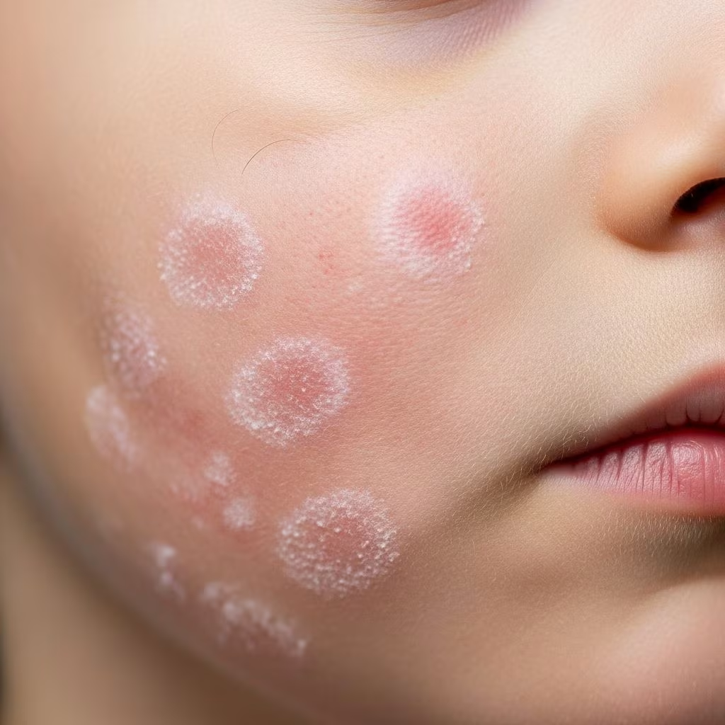
Pityriasis alba is a common, benign, self-limited superficial dermatitis, most commonly seen in children and adolescents. It presents as asymptomatic hypopigmented macules and patches with a characteristic fine scale that is accentuated by scratching or rubbing the skin surface. The term ‘pityriasis alba’ derives from ‘pityriasis’ (scaly) and ‘alba’ (white).
Pityriasis alba is more noticeable in summer when the surrounding skin tans, accentuating the hypopigmented patches. It is more obvious on dark skin types. It is very common, occurring in up to 80–100% of healthy children in some studies.

Who gets pityriasis alba?
Pityriasis alba affects children and adolescents, particularly those aged 6–12 years. It is equally common in males and females.
It is more prevalent in children with a personal or family history of atopic dermatitis, allergic rhinitis or asthma (ie, the atopic triad). Up to 80% of children with atopic eczema have pityriasis alba.
It occurs in all skin types but is more noticeable in those with Fitzpatrick skin types III–VI.
What causes pityriasis alba?
The exact cause of pityriasis alba is unknown.
The following factors have been implicated:
- Impaired stratum corneum barrier function in atopic individuals
- Chronic idiopathic inflammation with cytokine-mediated inhibition of melanogenesis
- Retained Malassezia spp lipophilic yeast cell fragments inducing inflammation
- Xerosis (dry skin)
- Sun exposure
What are the clinical features of pityriasis alba?
Pityriasis alba starts as red or pink patches with fine scaling, which are often itchy. Over time the erythema fades, leaving hypopigmented patches with a non-distinctive edge.
Primary lesions are round or oval macules or patches measuring 0.5–2 cm in diameter (but can be larger), with a characteristic fine scale that can be accentuated by rubbing the surface with a finger.
The face (cheeks) is most commonly affected (50%), followed by the upper arms, neck and trunk. Lesions on the face tend to be smaller and less scaly than those on the limbs.
Lesions may be single or multiple. There are usually 5–12 lesions, but there may be more than 20.
New lesions may appear over several months, but the condition tends to resolve spontaneously over 3–6 months (range 2 months–6 years).


Diagnosis
The diagnosis of pityriasis alba is based on the characteristic clinical appearance.
Dermoscopy shows a polymorphous ‘cornflake-like’ perifollicular scaling with absent pigment globules.
Differential diagnosis
Pityriasis alba can resemble other causes of acquired hypopigmentation. See Differential diagnosis of pityriasis alba table below.
| Condition | Key differentiating features |
|---|---|
| Postinflammatory hypopigmentation | History of preceding inflammation or trauma |
| Tinea corporis | Annular lesions with central clearing and raised border; +ve scraping/culture |
| Progression alba (Vitiligo) | Well-defined depigmented macules; chalky white; no scale |
| Pityriasis versicolor | Multiple fine scaling macules; confluence; scraping shows fungal hyphae/spores |
| Nummular dermatitis | Pruritic coin-shaped plaques; oozing/crusting |
| Psoriasis | Well-defined plaques with thick scale |
| Seborrhoeic dermatitis | Incorrect distribution; greasy scale |
| Contact dermatitis | History of exposure; geometric distribution |
| Idiopathic guttate hypomelanosis | Small round hypopigmented macules on sun-exposed shins/forearms of adults |
Investigations
No investigations are usually required to diagnose pityriasis alba.
Wood lamp examination shows patches to be off-white (not ‘glow’ as in vitiligo).
If diagnosis is uncertain, skin scrapings for microscopy and culture can exclude Malassezia (pityriasis versicolor), dermatophytes (tinea corporis) and yeast (Candida).
Skin biopsy is rarely required.
Histopathology
Histology of pityriasis alba shows:
- Mild compact orthokeratosis
- Reduced epidermal melanin and melanocyte numbers
- Perivascular lymphohistiocytic infiltrate
- Parakeratosis and spongiosis if early lesion
Late lesions show a normal epidermis with reduced and smaller melanosomes.

Management
No treatment is required for uncomplicated pityriasis alba as it is benign and self-limited.
Explanation and reassurance
Reassurance that the condition is harmless, non-contagious and will resolve in months is important.
Skin care
- Soap substitutes/emollients bd – td to treat xerosis and barrier dysfunction
- Sunscreen – hypopigmentation accentuated by tanning
Steroids
Mild topical corticosteroids eg, hydrocortisone 1% (apply nocte for 2 weeks then prn) can be used for itch.
Other
- Topical calcineurin inhibitors (tacrolimus 0.03%, pimecrolimus 1%) – reduce inflammation and repigment
- Tacrolimus 0.1% ointment – effective

Prognosis
Pityriasis alba is benign and self-limited. Most lesions resolve within 3–6 months but can take up to several years.
Recurrence is common, especially in atopic children.
It usually resolves by adulthood.
Prevention
Daily emollient use may prevent recurrences.
Related conditions
- Atopic dermatitis
- Postinflammatory hypopigmentation
- Vitiligo
- Pityriasis versicolor
Evidence-based therapy
| Intervention | Strength of evidence |
|---|---|
| Emollients | B |
| Hydrocortisone 1% | C |
| Topical calcineurin inhibitors | C |
Frequently asked questions
Is pityriasis alba permanent?
No. Pityriasis alba is self-limited and resolves spontaneously within months to years.
Is pityriasis alba eczema?
Pityriasis alba is a form of eczema/dermatitis that occurs in atopic children.
Does pityriasis alba go away?
Yes. It resolves spontaneously but may take several months.
Is pityriasis alba fungal?
No. Pityriasis alba is inflammatory, not infectious. Skin scrapings are negative.
Does pityriasis alba spread?
No. It is not contagious.
Can pityriasis alba be treated?
No treatment is necessary but emollients, hydrocortisone and tacrolimus ointment can help.
References
- Pityriasis Alba – StatPearls – NCBI Bookshelf — StatPearls Publishing. 2023. https://www.ncbi.nlm.nih.gov/books/NBK431061/
- Pityriasis alba: MedlinePlus Medical Encyclopedia — MedlinePlus, U.S. National Library of Medicine. 2024. https://medlineplus.gov/ency/article/001463.htm
- Pityriasis Alba: Causes, Symptoms, and Treatments – Healthline — Healthline Media. 2023. https://www.healthline.com/health/pityriasis-alba
- Pityriasis alba: Causes, symptoms, and treatment — Medical News Today. 2023. https://www.medicalnewstoday.com/articles/pityriasis-alba
- Pityriasis Alba: Symptoms, Causes & Treatment — Cleveland Clinic. 2024. https://my.clevelandclinic.org/health/diseases/pityriasis-alba
- All about pityriasis alba: causes, symptoms and treatments — Dexeryl. 2023. https://www.dexeryl.com/en/your-skin/pityriasis-alba



