
Introduction
Pityriasis lichenoides (PL) is an uncommon cutaneous rash of uncertain aetiology, characterised by a spectrum of inflammatory skin lesions. It encompasses two main forms at opposite ends of the disease spectrum: the acute form, known as pityriasis lichenoides et varioliformis acuta (PLEVA), and the chronic form, pityriasis lichenoides chronica (PLC). Many patients exhibit overlapping features of both, highlighting the condition’s variable presentation. PL is not contagious and primarily affects the skin, though rare systemic associations have been noted.
The condition typically manifests as recurrent crops of papules that evolve over time, often resolving with post-inflammatory changes such as hyperpigmentation or scarring. While generally benign, severe cases of PLEVA can lead to significant discomfort and cosmetic concerns. Understanding PL is crucial for accurate diagnosis and appropriate management, as it can mimic other dermatoses.
Demographics
Pityriasis lichenoides most commonly affects children and young adults, with PLEVA frequently observed in paediatric populations and young individuals under 30 years. It shows a slight male predominance, occurring more often in males than females. The condition can arise at any age, but peak incidence is in the first three decades of life. In children, it may present more aggressively as PLEVA, resembling chickenpox-like lesions.
Geographically, no specific predilection is noted, though reports suggest higher recognition in temperate climates where dermatological surveillance is robust. Ethnic variations are minimal, but lesion colour may differ: erythematous on light skin, violaceous or brown on darker skin tones. Incidence is rare, estimated at less than 1% of dermatology referrals, underscoring its uncommon nature.
Causes
The exact cause of pityriasis lichenoides remains unknown, but several hypotheses exist based on histopathological and clinical observations. It is postulated as a hypersensitivity reaction to foreign antigens, such as infections (bacterial or viral) or medications. The childhood form particularly suggests a reactive process to pathogens, though no specific trigger is consistently identified.
- Infectious triggers: Associations with Epstein-Barr virus, adenovirus, parvovirus, and bacterial toxins have been reported, though causality is unproven.
- Immune dysregulation: PL features a lymphocytic infiltrate, hinting at T-cell mediated immunity, potentially linking to cutaneous T-cell lymphoma in rare malignant transformations.
- Other factors: Vaccinations, drugs, or environmental antigens may precipitate episodes, but evidence is anecdotal.
Importantly, PL is not hereditary nor infectious, dispelling common misconceptions. Recent studies explore type 2 inflammation in refractory cases, with elevated IgE and eosinophils in some patients.
Clinical Features
Clinical presentation varies between acute and chronic forms, often overlapping in a single patient.
Pityriasis Lichenoides et Varioliformis Acuta (PLEVA)
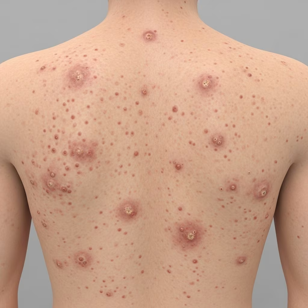
PLEVA presents abruptly with a rapidly progressive rash of crops of erythematous papules (2–10 mm), which evolve into vesicles, pustules, or necrotic centres. Lesions are often asymptomatic initially but may become pruritic or painful. They appear on the trunk, proximal limbs, and flexures, resembling chickenpox or varioliform eruptions.
- New lesions: Pink/red papules with central necrosis or ulceration.
- Evolution: Crusting, haemorrhagic scabs heal over 2–4 weeks, often leaving scars or hypopigmentation.
- Skin tone variation: Violaceous-brown on darker skin.
Mucosal involvement (oral, genital) occurs rarely.
Pityriasis Lichenoides Chronica (PLC)
PLC develops more insidiously over days to weeks with flatter, reddish-brown papules (3–8 mm) featuring adherent scale. Lesions are mildly pruritic and resolve slowly (4–12 weeks) with hyperpigmentation.
- Common sites: Trunk, thighs, flexural areas.
- Chronicity: Waxing/waning for months to years; PLEVA may transition to PLC.
Patients often have polymorphic lesions: acute crops amid chronic patches.
Complications
Most cases are self-limited, but complications include:
- Scarring: Atrophic or hypertrophic scars from necrotic PLEVA lesions, especially in children.
- Hyperpigmentation/hypopigmentation: Post-inflammatory changes, more noticeable in darker skin.
- Secondary infection: Rare, from excoriation of ulcerated lesions.
- Rare severe forms: Febrile ulceronecrotic Mucha-Habermann disease (FUMHD), a PLEVA variant with systemic symptoms, high fever, and potential mortality.
- Malignant potential: Very rare progression to cutaneous T-cell lymphoma.
Diagnosis
Diagnosis is primarily clinical, supported by skin biopsy. Key histopathological features include:
- Interface dermatitis with vacuolar degeneration.
- Dense lymphocytic infiltrate in dermis/epidermis.
- Exocytosis, parakeratosis, and necrotic keratinocytes (more in PLEVA).
No specific laboratory tests; rule out infections or lymphoma if atypical.
Differential Diagnoses
| Condition | Key Distinguishing Features |
|---|---|
| Chickenpox/VZV | Systemic symptoms, vesicles in crops, positive Tzanck smear. |
| Pityriasis rosea | Herald patch, Christmas-tree distribution, self-resolves 6–8 weeks. |
| Guttate psoriasis | Silver scale, Auspitz sign, streptococcal trigger. |
| Lymphomatoid papulosis | Larger lesions, CD30+ cells on biopsy, lymphoma association. |
| Drug eruption | History of new medication, eosinophilia. |
Treatment
No curative treatment exists; management targets symptoms and hastens resolution. Observation suffices for mild cases.
First-Line Therapies
- Topical glucocorticoids: Potent steroids (e.g., clobetasol) for inflammation/pruritus.
- Topical tacrolimus: Calcineurin inhibitor for steroid-sparing.
- Oral antibiotics: Erythromycin (children) or tetracyclines (adults) for 1–3 months; anti-inflammatory effect.
- Natural sunlight: Helpful adjunct.
Second-Line/Phototherapy
- Narrow-band UVB (NB-UVB): Most effective (47% response); weekly sessions.
- PUVA: Psoralen + UVA for refractory cases, higher side-effect risk.
Advanced Therapies
- Immunosuppressants: Methotrexate, cyclosporine for severe/refractory disease.
- Biologics: Emerging; stapokibart (IL-4/13 inhibitor) showed complete resolution in a case with type 2 inflammation after 12 weeks; dupilumab reported similarly.
Treatment response varies; phototherapy tops efficacy per cohorts. Monitor for side effects, especially in children.
Outcome
PLEVA typically resolves in weeks to months, often with scarring. PLC persists months to years, waxing/waning; 50–80% remit within 1–3 years. Recurrences common but severity decreases over time. Prognosis excellent; rare progression to malignancy. Patient education on self-limited nature aids coping.
Frequently Asked Questions (FAQs)
Q: Is pityriasis lichenoides contagious?
A: No, PL is not infectious and cannot be spread to others.
Q: Does it go away on its own?
A: Yes, most cases self-resolve within months to years without treatment.
Q: What is the best treatment for children?
A: Erythromycin, topical steroids, and phototherapy are preferred; avoid systemic immunosuppressants initially.
Q: Can it scar?
A: PLEVA often leaves small scars from necrotic lesions; PLC causes pigmentation changes.
Q: Is it related to cancer?
A: Rare association with lymphoma; monitor atypical/persistent cases with biopsy.
References
- Pityriasis Lichenoides — Merck Manuals Professional Edition. 2024. https://www.merckmanuals.com/professional/dermatologic-disorders/psoriasis-and-other-papulosquamous-disorders/pityriasis-lichenoides
- Successful treatment of pityriasis lichenoides et varioliformis acuta with stapokibart — Frontiers in Medicine. 2025. https://www.frontiersin.org/journals/medicine/articles/10.3389/fmed.2025.1615108/full
- Pityriasis Lichenoides Patient Information — Skin Health Info (BAD). 2016 (reviewed 2018). https://www.skinhealthinfo.org.uk/wp-content/uploads/2018/11/Pityriasis-lichenoides-Update-September-2016-lay-reviewed-July-20163.pdf
- Pityriasis Lichenoides Chronica in Children — Children’s Health. 2025. https://www.childrens.com/specialties-services/conditions/pityriasis-lichenoides
- Pityriasis Lichenoides — DermNet NZ. 2025. https://dermnetnz.org/topics/pityriasis-lichenoides



