
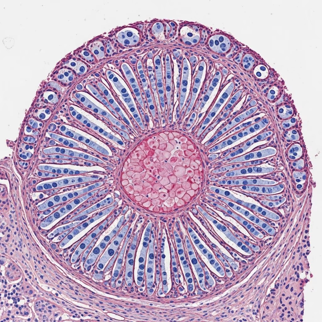
Histology of rheumatoid nodule
In rheumatoid nodules, low-power microscopic examination reveals a distinctive granulomatous tissue reaction pattern characterized by well-circumscribed lesions within the dermis, often extending deeply into subcutaneous tissues (Figure 1).
Central to the pathology are well-formed necrobiotic granulomas, consisting of large areas of eosinophilic necrosis with fibrinoid material deposition. These foci are sharply demarcated and surrounded by a characteristic palisading arrangement of histiocytes—radially oriented macrophages forming a fence-like border around the necrotic core (Figure 2).
The palisade is accompanied by a mixed inflammatory infiltrate including lymphocytes, plasma cells, multinucleated giant cells (often Langhans-type), and occasional eosinophils. Perivascular lymphocytic cuffing is common, reflecting the vasculocentric nature of the process (Figure 3).
Lesions frequently involve the deep dermis and subcutis, with extension around adnexal structures or along fascial planes. Fibrosis may develop in older nodules, partially replacing the granulomatous reaction.
Cytology description
- Necrobiotic focus: Homogeneous, eosinophilic, fibrinoid necrosis without nuclear debris; sharply demarcated.
- Palisading histiocytes: Elongated nuclei oriented perpendicular to the necrotic border; abundant pale cytoplasm.
- Multinucleated giant cells: Scattered throughout the palisade; horseshoe or ring-shaped nuclei.
- Inflammatory cells: Lymphocytes (predominant), plasma cells, rare eosinophils; perivascular accentuation.
- Stroma: Delicate vascularity with endothelial swelling; early fibrosis in chronic lesions.
- No vasculitis: Absent endothelial damage or fibrin thrombi distinguishes from true vasculitides.
Histological images
- Figure 1: Scanning magnification showing granulomatous pattern with central necrosis.
- Figure 2: Necrobiotic granuloma with palisading histiocytes.
- Figure 3: High-power view of mixed infiltrate and giant cells.
These images demonstrate the classic triad: central fibrinoid necrosis, palisading granuloma, and mixed inflammation.
Pathogenesis
Rheumatoid nodules represent an exaggerated Type IV hypersensitivity reaction driven by rheumatoid arthritis (RA) autoimmunity. Trauma to extensor surfaces triggers microvasculature injury, releasing chemotactic factors that recruit T-cells, macrophages, and B-cells.
Key pathogenetic features include:
- Complement activation: Deposits rheumatoid factor (RF)-immune complexes along vessels.
- Cytokine cascade: TNF-α, IL-1, IL-6 drive histiocyte activation and granuloma formation.
- Fibrin deposition: Extravasated plasma proteins undergo denaturation, forming the eosinophilic necrotic core.
- Genetic predisposition: Strong association with HLA-DR4 and high-titer RF/ACPA positivity.
Nodules develop in 20-40% of RA patients, particularly seropositive males with severe disease, and correlate with accelerated atherosclerosis and pulmonary involvement.
Clinical features
Rheumatoid nodules are the most common extra-articular manifestation of RA, affecting 35-40% of patients, especially RF-positive Caucasian males.
| Feature | Description |
|---|---|
| Location | Extensor surfaces (elbows, forearms, knuckles, heels); pressure points |
| Size | 2 mm to 5 cm; firm, mobile subcutaneous lumps |
| Symptoms | Asymptomatic; rarely painful unless ulcerated/infected |
| Associations | Severe RA, RF+, HLA-DR4, smoking, methotrexate use |
| Complications | Ulceration, infection, fistula formation, nerve compression |
Subcutaneous nodules appear within 2 years in 5% of RA cases, rising to 30% lifetime prevalence.
Diagnosis
Diagnosis combines clinical context (RA history, extensor nodules) with histology. Serology (RF, ACPA) and imaging (joint erosions) support but are not required for nodule confirmation.
- Gold standard: Biopsy showing palisading necrobiotic granuloma.
- Serology: 90% RF-positive; high titers predict nodule formation.
- Imaging: Ultrasound shows hypoechoic masses; MRI delineates deep extension.
Differential diagnosis
Rheumatoid nodules must be distinguished from other palisading granulomas:
| Condition | Key Distinguishing Features |
|---|---|
| Granuloma annulare | Mucin deposition (Alcian blue+); smaller necrobiosis; no fibrinoid necrosis; superficial dermis |
| Necrobiosis lipoidica | Layered necrobiosis; plasma cells; atrophic epidermis; diabetes association; mucin absent |
| Gouty tophus | Amorphous urate crystals (polarized light); fewer inflammatory cells |
| Sarcoidosis | Confluent naked granulomas; no necrosis; asteroid bodies |
| Infectious granuloma | Caseating necrosis; organisms (AFB, GMS); suppuration |
Clinical history (RA presence) is often diagnostic; special stains (mucin, crystals, organisms) resolve mimics.
Rheumatoid nodulosis
A benign variant in males 30-50 years with recurrent, self-limiting nodules but minimal arthritis. Features intraosseous cysts, RF+, and occasional progression to classic RA.
Complications
- Skin breakdown → secondary infection (Staph, Pseudomonas)
- Deep nodules → fistula tracts, osteomyelitis
- Cardiovascular: Independent risk factor for MI, stroke
- Pulmonary: Pleural nodules, interstitial disease
Treatment
Nodules are managed conservatively unless symptomatic:
- Intralesional steroids: Triamcinolone 10-40 mg/mL; 70-90% response rate.
- Surgery: Excision for persistent/ulcerated lesions.
- DMARDs: Methotrexate may paradoxically increase nodules; switch to biologics (anti-TNF, rituximab).
- Observation: 20-30% spontaneous regression.
Frequently Asked Questions (FAQs)
Q: What is the hallmark histological feature of rheumatoid nodules?
A: Palisading necrobiotic granulomas with central fibrinoid necrosis surrounded by histiocytes, lymphocytes, and giant cells.
Q: Are rheumatoid nodules painful?
A: Typically asymptomatic, but may cause discomfort if infected, ulcerated, or compressing nerves.
Q: How common are nodules in rheumatoid arthritis?
A: 20-40% lifetime prevalence, higher in seropositive males with severe disease.
Q: Can nodules appear before joint symptoms?
A: Yes, in rheumatoid nodulosis—multiple nodules with minimal arthritis and good prognosis.
Q: What stains help differentiate from granuloma annulare?
A: Mucin stains (Alcian blue, colloidal iron) negative in rheumatoid nodules but positive in GA.
References
- Rheumatoid nodule – Wikipedia — Wikipedia contributors. 2024-01-15. https://en.wikipedia.org/wiki/Rheumatoid_nodule
- Recognising skin manifestations of rheumatological disease — Royal Australian College of General Practitioners (RACGP). 2021-12-01. https://www1.racgp.org.au/ajgp/2021/december/skin-manifestations-of-rheumatological-disease
- Rheumatoid nodule pathology — DermNet NZ. 2024. https://dermnetnz.org/topics/rheumatoid-nodule-pathology
- Rheumatoid arthritis and the skin — DermNet NZ. 2024. https://dermnetnz.org/topics/rheumatoid-arthritis-and-the-skin
- Rheumatoid nodule pathology image — DermNet NZ. 2024. https://dermnetnz.org/imagedetail/18351-rheumatoid-nodule-pathology
- Granuloma annulare pathology — DermNet NZ. 2024. https://dermnetnz.org/topics/granuloma-annulare-pathology



