
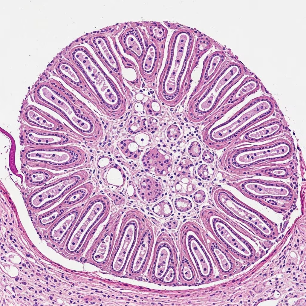
Schwannoma, also known as neurilemmoma, neurolemmoma, or Schwann cell tumour, is a benign peripheral nerve sheath tumour composed predominantly of Schwann cells. These cells normally produce the insulating myelin sheath that covers peripheral nerves, ensuring efficient nerve signal transmission. Schwannomas typically arise from sensory nerves and are the most common neurogenic tumours encountered in clinical practice.
Introduction
Schwannomas represent approximately 5% of benign soft tissue tumours in adults and 2% in children, with cutaneous manifestations accounting for a notable subset. They most frequently occur in the head and neck region (25-45% of cases), flexor surfaces of extremities, and trunk, though lower limb involvement, as in rare atypical presentations, comprises only about 1% of cases. Clinically, these tumours present as solitary, slowly growing, painless, firm subcutaneous nodules ranging from 0.25 cm to 3 cm in diameter, though larger lesions up to 7 cm have been documented.
While 90% occur as isolated lesions, associations exist with neurofibromatosis type 2 (3%), schwannomatosis (2%), and central nervous system tumours like meningiomas (5%). Pain or paresthesia may arise from compression of adjacent nerve structures. Histologically, schwannomas are hallmark encapsulated lesions, distinguishing them from other nerve sheath tumours.
Clinical Features
- Solitary nodule: Firm, mobile, subcutaneous mass with intact overlying epidermis.
- Size: Typically 1-3 cm; slow growth over years, occasionally rapid enlargement.
- Symptoms: Usually asymptomatic; tenderness or Tinel’s sign (radiating paresthesia on percussion) in 30-50% due to nerve involvement.
- Locations: Head/neck (commonest), extremities, trunk; rare on lower limbs.
- Associations: NF2, schwannomatosis, Carney complex (melanotic variant).
Imaging such as ultrasound reveals a well-defined, hyperechoic mass without central vascularity, often mimicking lipoma. MRI shows T2 hyperintensity with peripheral enhancement, confirming encapsulation.
Histopathology
Microscopic examination reveals a well-circumscribed, encapsulated lesion in the dermis or subcutis beneath uninterrupted epidermis. The tumour exhibits biphasic architecture with alternating areas of varying cellularity.
Antoni A Areas
These are hypercellular regions comprising haphazardly arranged bland spindle cells with elongated, wavy nuclei and eosinophilic cytoplasm. Cells form short fascicles or whorls. Characteristic Verocay bodies—parallel palisades of nuclei flanking acellular eosinophilic hyalinised collagen cores—are diagnostic (present in 10-20% of cases).
Antoni B Areas
Hypocellular zones feature a loose, oedematous, mucinous stroma with fibrillar collagen, hypocellular spindle cells, and prominent, often hyalinised blood vessels surrounded by dense sclerosis. Myxoid change, haemorrhage, and hemosiderin deposition are common.
Other Features
- Encapsulation: Thin perineurial capsule (EMA-positive).
- Associated nerve: Eccentric peripheral nerve often identifiable.
- Ancient change: Degenerative atypia—pleomorphic, hyperchromatic nuclei, multinucleation—without mitoses or necrosis; represents reactive phenomenon in long-standing lesions.
- Vascular changes: Thickened, hyalinised walls with fibrin thrombi and haemorrhage.
| Feature | Description | Significance |
|---|---|---|
| Antoni A | Cellular, palisading, Verocay bodies | Diagnostic hallmark |
| Antoni B | Loose myxoid stroma, vessels | Contrasts with A areas |
| Encapsulation | Perineurial sheath | Benign behaviour |
| Ancient atypia | Pleomorphic nuclei | Non-malignant |
Cytology
Fine-needle aspiration yields cellular smears of spindle cells in metachromatic stroma, with nuclear grooves and Verocay-like palisading. However, core biopsy or excision is preferred for definitive diagnosis due to sampling limitations.
Immunohistochemistry
Schwannomas show strong, diffuse S100 positivity (nuclear and cytoplasmic), confirming Schwannian differentiation. SOX10 is also positive. Capsule expresses EMA. Neurofilament highlights entrapped axons peripherally, but axons are absent within the tumour, unlike neurofibroma.
- S100: Strong positive
- SOX10: Positive
- EMA: Capsule positive
- Neurofilament: Peripheral nerves
- CD34: Negative (vs. DFSP)
Differential Diagnosis
Key mimics include:
| Tumour | Key Distinguishing Features |
|---|---|
| Neurofibroma | Lacks encapsulation, Antoni patterns; axons course through (neurofilament+); CD34+, S100 weaker |
| Plexiform neurofibroma | Plexiform growth, NF1 association; no Verocay bodies |
| Perineurioma | EMA+, S100-; slender processes, collagen rosettes |
| DFSP | CD34+, S100-; storiform, infiltrative, no encapsulation |
| MPNST | Infiltrative, high mitoses, necrosis; focal S100; rapid growth |
| Cellular schwannoma | Predominantly Antoni A-like, no B areas; mitoses low |
| Palisaded encapsulated neuroma | Axons throughout (neurofilament+), less myxoid |
Variants
- Cellular schwannoma: Hypercellular, simulates MPNST; lacks necrosis/mitoses.
- Plexiform schwannoma: Rope-like, childhood; malignant potential rare.
- Melanotic (psammomatous): Pigmented, Carney complex; psammoma bodies.
- Ancient schwannoma: Degenerative atypia.
Treatment and Prognosis
Complete surgical excision or enucleation is curative, with <1% recurrence. Nerve function preservation is prioritised; paresthesia may persist post-excision. Malignant transformation is exceedingly rare.
Frequently Asked Questions (FAQs)
Q: What is the hallmark histological feature of schwannoma?
A: Biphasic Antoni A (cellular with Verocay bodies) and Antoni B (loose myxoid) areas within an encapsulated nodule.
Q: How does schwannoma differ from neurofibroma pathologically?
A: Schwannoma is encapsulated, lacks intratumoural axons, shows Antoni patterns; neurofibroma is unencapsulated with axons throughout.
Q: Is S100 staining diagnostic for schwannoma?
A: Strong S100 positivity supports Schwannian origin but requires architecture for diagnosis; differentials like MPNST may be focal S100+.
Q: Can schwannomas be malignant?
A: Conventional schwannomas are benign; rare progression in plexiform/cellular variants or NF-associated cases.
Q: What is ancient change in schwannoma?
A: Reactive atypia with pleomorphic nuclei in longstanding tumours; no mitoses/necrosis, benign behaviour.
References
- Schwannoma pathology — DermNet NZ. 2025. https://dermnetnz.org/topics/schwannoma-pathology
- The pathobiologic spectrum of Schwannomas — PubMed (Woodruff JM et al.). 2003-06-01. https://pubmed.ncbi.nlm.nih.gov/12792904/
- Cutaneous schwannoma: an atypical presentation — PMC (Alkhalifah A et al.). 2017-05-01. https://pmc.ncbi.nlm.nih.gov/articles/PMC5514603/
- Palisaded and encapsulated neuroma pathology — DermNet NZ. 2025. https://dermnetnz.org/topics/palisaded-and-encapsulated-neuroma-pathology



