Sneddon syndrome is a rare, slowly progressive, non-inflammatory thrombotic vasculopathy characterized by the triad of livedo reticularis, recurrent cerebrovascular ischaemic events, and early-onset dementia.
It typically affects young to middle-aged women and involves small- to medium-sized arteries of the skin, brain, heart, kidneys, and eyes. Livedo reticularis—a mottled, bluish-purple, fishnet-like skin discoloration—often precedes neurological symptoms by years, serving as a critical diagnostic clue.
What is Sneddon syndrome?
Sneddon syndrome (SS) is defined by the association of generalized livedo reticularis (or livedo racemosa) with multiple cerebrovascular episodes, including transient ischemic attacks (TIAs) and ischemic strokes. First described by Ian Bruce Sneddon in 1965, it affects approximately 0.1 per 100,000 individuals, predominantly women (female-to-male ratio 4:1), with onset typically between ages 20–42 years.
The underlying pathology involves non-inflammatory occlusion of arterioles and small arteries by intimal proliferation and fibrosis, leading to endothelial proliferation without significant inflammation or thrombosis in many cases. This distinguishes SS from inflammatory vasculitides like polyarteritis nodosa.
SS progresses in stages: a prodromal phase with headaches and dizziness (up to 9 years), followed by TIAs/strokes, and culminating in cognitive decline and dementia. Multi-organ involvement includes the heart (valvular disease), kidneys (renal impairment), and eyes (retinal vascular occlusions).
Who gets Sneddon syndrome (Epidemiology)?
- Primarily young adults aged 20–50 years, with mean onset at 32 years.
- Strong female predominance (70–80% of cases).
- Rare overall incidence: <4 cases per million population annually.
- No strong racial or geographic predilection reported, though cases are documented worldwide.
- Up to 10–20% of patients with idiopathic livedo reticularis may develop SS.
Clinical features
Stage I: Prodromal phase
Headaches (often migrainous, triggered by cold, stress, or menses) and vertigo/dizziness precede skin or focal neurological changes by 1–9 years in 50–70% of patients. These are nonspecific and often lead to delayed diagnosis.
Stage II: Stroke-prone phase
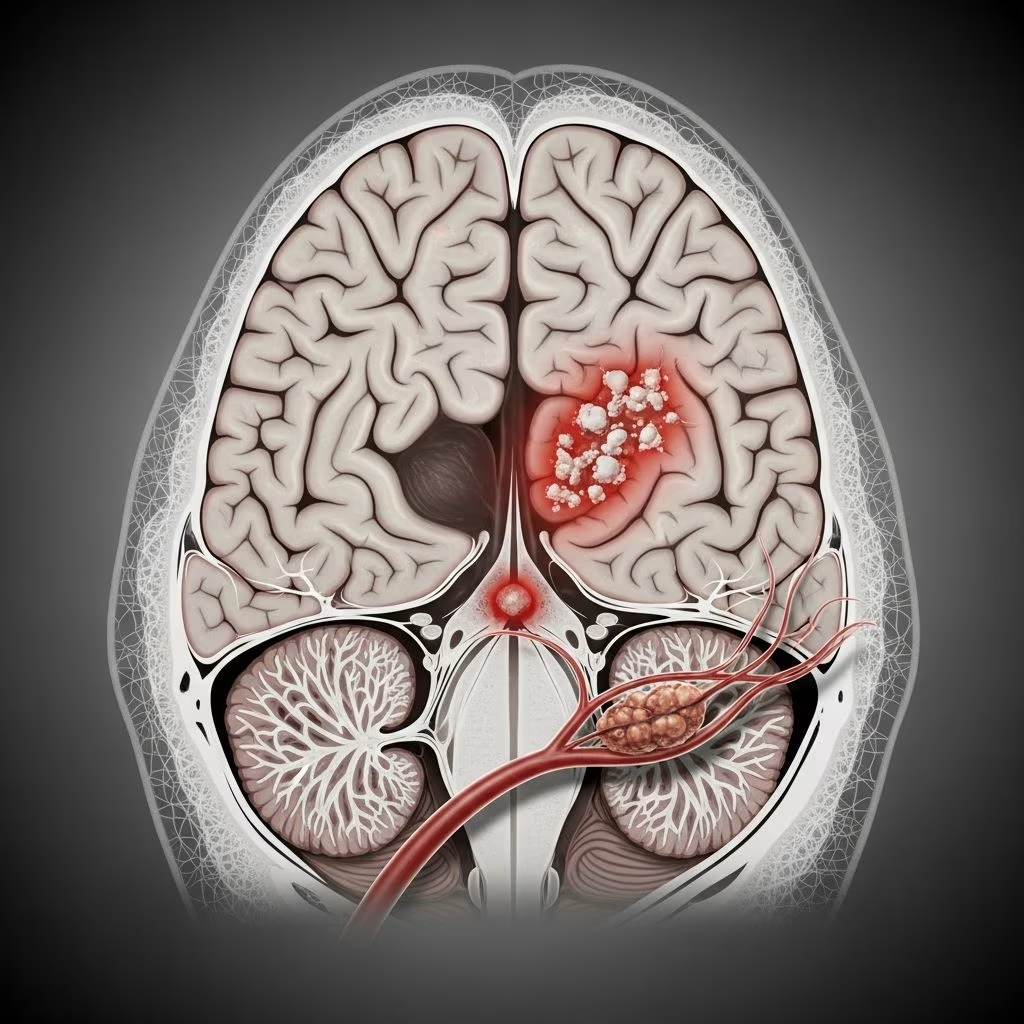
Recurrent TIAs (70–80%) and ischemic strokes (55–75%), predominantly in middle cerebral artery (MCA) territory, causing hemiparesis (most common), aphasia, hemianopia, or sensory deficits. Strokes are cortical/subcortical, with TIAs recurring weekly to yearly.
Stage III: Cognitive decline
Progressive dementia in 40–75% by 5–13 years post-onset, mimicking Alzheimer’s with deficits in memory, attention, visuospatial skills, and executive function. Psychiatric features include depression (30%), psychosis, and suicide risk. Seizures occur in 15–20%, often post-stroke.
Skin features
Generalized livedo reticularis/racemosa (broken, irregular net-like purple discoloration) on trunk, buttocks, thighs, and arms, exacerbated by cold. Present in >99% of cases, often antedating strokes by >10 years. Skin biopsies show endothelial proliferation and arteriolar occlusion.
Other features
- Cardiac: Valvular stenosis/vegetations (20–30%), ischemic heart disease, murmurs.
- Renal: Hypertension (35–50%), proteinuria, renal infarcts (10–15%).
- Ocular: Retinal artery/vein occlusions, optic neuropathy, hemianopia (15–25%). Central retinal vein occlusion is most common.
- Musculoskeletal: Rare myalgia, arthralgia, chorea.
Complications
Major complications include recurrent strokes (50% risk despite antiplatelets), vascular dementia (poor prognosis), myocardial infarction, renal failure, and blindness from retinal ischemia. Mortality is 10–15% at 10 years, mainly from stroke/heart disease. Pregnancy increases stroke risk.
Diagnosis
Diagnosis is clinical, requiring livedo reticularis + >1 ischemic stroke/TIA without other cause. Median diagnostic delay: 2–6 years.
| Diagnostic Criteria | Essential Features |
|---|---|
| Clinical | Livedo reticularis + cerebrovascular ischaemia (TIA/stroke) |
| Exclusion | No systemic vasculitis, coagulopathy, or endocarditis |
| Supportive | Antiphospholipid antibodies (20–40%), hypertension |
Investigations
- Skin biopsy: Endarteritis obliterans (noninflammatory intimal fibrosis).
- Brain MRI: Multifocal infarcts, white matter lesions.
- Echocardiogram: Valvular abnormalities.
- Labs: Normal ESR/CRP; antiphospholipid antibodies (lupus anticoagulant, anticardiolipin) in 20–50%; exclude coagulopathies.
- Angiography: Arterial stenoses/occlusions.
Differential diagnosis
| Condition | Key Distinguishing Features |
|---|---|
| Antiphospholipid syndrome | Thrombosis + aPL antibodies; less livedo; systemic involvement |
| Painful ulcers; legs only; no strokes | |
| Primary CNS vasculitis | Inflammatory markers elevated; biopsy shows inflammation |
| Atheroembolic disease | Older age; cholesterol emboli on biopsy |
| Polyarteritis nodosa | Necrotizing inflammation; aneurysms on angio |
Management
No randomized trials; management is empirical, focusing on stroke prevention and symptom control.
- Antiplatelet therapy: Aspirin 75–325 mg daily (first-line); reduces recurrence by 50–70%.
- Anticoagulation: Warfarin (INR 2–3) if aPL-positive, venous thrombosis, or cardioembolic source.
- Statins: For hyperlipidemia/arterial disease.
- Antihypertensives: Target BP <130/80 mmHg.
- Symptomatics: Migraine prophylaxis (topiramate), antidepressants, antiepileptics.
- Experimental: IVIG, statins, anti-TNF (anecdotal).
Avoid estrogens/OCPs due to thrombotic risk. Multidisciplinary care (neurology, dermatology, rheumatology) essential.
Prognosis
Guarded: 40–50% stroke-free at 10 years with antiplatelets; 45–75% develop dementia. Poor visuospatial/attention outcomes. Annual stroke risk 8–15%; mortality from recurrent CVA.
Prevention
- Smoking cessation, healthy lifestyle.
- Early recognition of livedo in young patients.
- Strict adherence to antiplatelet/anticoagulant therapy.
Patient education
Counsel on cold avoidance (exacerbates livedo), stroke warning signs (FAST: Face/Arm/Speech/Time), medication compliance, and pregnancy risks. Regular neurocognitive monitoring recommended.
Frequently Asked Questions
Is Sneddon syndrome hereditary?
No established genetic cause; rare familial cases reported. Not routinely inherited.
Does livedo reticularis always mean Sneddon syndrome?
No; common in cold exposure, coagulopathies. Strokes + generalized livedo diagnostic.
Can Sneddon syndrome be cured?
No cure; treatable with antiplatelets to prevent progression.
Is pregnancy safe with Sneddon syndrome?
High stroke risk; requires anticoagulation/heparin bridging.
What is the life expectancy?
Reduced; 10-year survival ~85%, limited by strokes/dementia.
References
- Sneddon Syndrome – Symptoms, Causes, Treatment — National Organization for Rare Disorders (NORD). 2023. https://rarediseases.org/rare-diseases/sneddon-syndrome/
- Comprehensive insights of Sneddon syndrome: A clinical perspective — SAGE Journals. 2024-10-17. https://journals.sagepub.com/doi/10.1177/11795735241308767
- Sneddon Syndrome: A Comprehensive Overview — PubMed/NCBI. 2019-06-01. https://pubmed.ncbi.nlm.nih.gov/31160219/
- Sneddon’s syndrome: a comprehensive review of the literature — PMC/NCBI. 2015-01-20. https://pmc.ncbi.nlm.nih.gov/articles/PMC4302600/
- Sneddon’s Syndrome: Symptoms and Treatment — Patient.info. 2024. https://patient.info/doctor/multisystem-diseases/sneddons-syndrome