
Author: Dr. Priya Singh, Dermatopathologist
Revised: January 2026
What is spindle cell lipoma?
Spindle cell lipoma is a distinctive, benign adipocytic neoplasm characterized by a mixture of mature adipocytes, bland spindle cells, and thick ropey collagen bundles. First described by Enzinger and Harvey in 1975, it represents approximately 1.5% of all lipomas and typically arises in the subcutaneous tissue.
This tumor is composed of variable proportions of fat cells and spindle cells embedded in a myxoid or fibrous stroma. Unlike conventional lipomas, which consist primarily of mature adipocytes, spindle cell lipomas feature a biphasic pattern with spindle cell proliferation that can mimic more aggressive lesions.
The lesion grows slowly, measures 1-5 cm on average (range 1-14 cm), and is usually solitary. Rare cases of multiple or familial occurrences have been documented.
Terminology
- Spindle cell lipoma (SCL)
- Pleomorphic lipoma (when floret-type giant cells are present; considered part of the same spectrum)
- Shaw area lipoma (due to predilection for posterior neck/shoulder region)
Epidemiology
Spindle cell lipomas predominantly affect males (male-to-female ratio approximately 10:1), with peak incidence in the 45-65 year age group.
In females, lesions tend to occur at younger ages and in atypical locations outside the classic “shawl” distribution (posterior neck, shoulder, upper back).
| Demographic | Characteristics |
|---|---|
| Gender | Male predominance (90%+ cases) |
| Age | 45-65 years (peak 6th decade) |
| Incidence | 1.5% of all lipomas |
Clinical features
Location
Classic sites (95% of cases):
- Posterior neck (most common)
- Shoulder
- Upper back (“shawl zone”)
Atypical sites (5%):
- Anterior neck, face, extremities, oral cavity, mediastinum, breast
Presentation
- Slow-growing, painless subcutaneous mass
- Firm or rubbery consistency (due to collagen content)
- Mobile over underlying structures
- Well-circumscribed, oval/discoid shape
- Skin overlying lesion typically normal
Grossly, cut surface appears yellow-white with fibrous bands.
Pathology
Histology
Spindle cell lipomas exhibit a biphasic pattern composed of:
- Mature adipocytes (variable proportion, 10-90%)
- Bland spindle cells with oval nuclei, scant cytoplasm
- Ropey collagen bundles (characteristic “rope-like” thick fibers)
- Myxoid or fibrous stroma
Architectural patterns:
- Short fascicles of spindle cells (parallel or storiform)
- Palisading of nuclei
- Mast cell aggregates (common)
- Biphasic areas with adipocytes and spindle cells
No atypia, mitoses, or necrosis. In pleomorphic lipoma variant, floret-type giant cells may be present.
Cytology
- Mixture of adipocytes and oval/spindle cells
- Metachromatic stroma (myxoid)
- Thick collagen fibers
- Mast cells
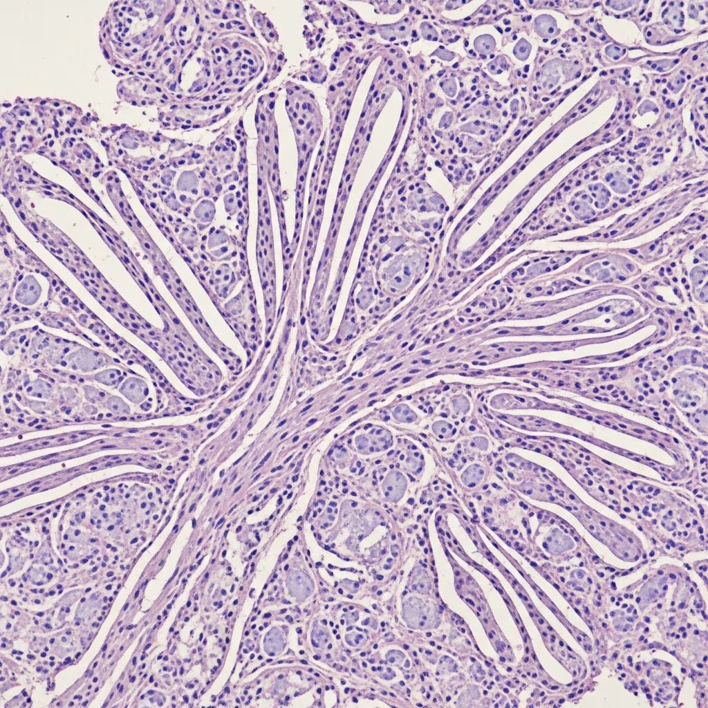
Histopathology images
(Description: Low-power view shows well-circumscribed lesion with adipocytes, spindle cell fascicles, and collagen bundles. High-power demonstrates bland spindle cells without atypia.)
Histopathology — differential diagnosis
| Diagnosis | Key distinguishing features |
|---|---|
| Well-differentiated liposarcoma | ATFIRMD adipocytes, lipoblasts, MDM2 amplification, no ropey collagen, deeper location |
| Myofibroma | More cellular, nodular pattern, SMA+, factor XIIIa+ |
| Neurofibroma | S100+, wavy nuclei, less fat, mucin-rich |
| Solitary fibrous tumor | Patternless pattern, STAT6+, more hemangiopericytoma-like vessels |
| Low-grade fibromyxoid sarcoma | ARC/collagen rosettes, MUC4+, atypia |
| Cellular schwannoma | Strong S100+, Verocay bodies, ancient change |
Immunohistochemistry
| Marker | Staining pattern | Notes |
|---|---|---|
| CD34 | Positive (spindle cells) | Diffuse, strong |
| S100 | Negative | Crucial negative stain |
| RB1 | Loss of expression | Diagnostic (complete absence) |
| Desmin | Negative | |
| SOX10 | Negative | Distinguishes from neural tumors |
| STAT6 | Negative | Excludes solitary fibrous tumor |
| MDM2 | Negative | Excludes liposarcoma |
| MUC4 | Negative | Excludes LGFMS |
Genetics
Characteristic cytogenetic abnormalities:
- Loss of 13q14 (RB1 locus) — 80-90% of cases
- Loss of 16q — common
- Loss of 12q — occasional
No MDM2 or CDK4 amplification (distinguishes from liposarcoma).
Prognosis
- Excellent — benign neoplasm with no metastatic potential
- No reports of malignant transformation
- Recurrence rare (<5%) after complete excision
Treatment
- Simple excision — curative treatment
- Marginal excision sufficient (well-circumscribed)
- No adjuvant therapy required
- Observation acceptable for asymptomatic small lesions
Imaging
Non-specific findings:
- Well-circumscribed subcutaneous mass
- Variable fat content (depending on adipocyte proportion)
- Heterogeneous enhancement on MRI
- No calcification
Frequently asked questions
Q: Is spindle cell lipoma cancerous?
A: No. Spindle cell lipoma is a benign tumor with no malignant potential. Complete surgical excision is curative.
Q: How can spindle cell lipoma be distinguished from liposarcoma?
A: Key features include superficial location, ropey collagen, CD34+/S100-, RB1 loss, no MDM2 amplification, and absence of lipoblasts/atypia.
Q: Why do spindle cell lipomas predominantly affect men?
A: Androgen receptors have been identified in these tumors, potentially explaining the strong male predilection.
Q: What is the treatment for spindle cell lipoma?
A: Simple surgical excision is the treatment of choice and is curative. Recurrence is exceptionally rare.
Q: Can spindle cell lipomas occur in unusual locations?
A: Yes, though rare. Reported sites include oral cavity, mediastinum, breast, and extremities, particularly in women.
References
- Spindle-cell Lipoma of the Skin — PubMed/NCBI. 1995-11-01. https://pubmed.ncbi.nlm.nih.gov/8599462/
- Spindle Cell Lipoma: A Rare, Misunderstood Entity — PMC/NIH. 2017-08-07. https://pmc.ncbi.nlm.nih.gov/articles/PMC5556811/
- Fat Tissue Abnormalities – Spindle Cell Lipoma — Perridermatology. 2023. https://perridermatology.com/dr-perris-blog/fat-tissue-abnormalities-spindle-cell-lipoma/
- Spindle Cell Lipoma and Pleomorphic Lipoma: An Update and Review — Cancer Diagnosis & Prognosis. 2022. https://www.cancerdiagnosisprognosis.org/article/214/spindle-cell-lipoma-and-pleomorphic-lipoma-an-update-and-review
- Spindle Cell Lipoma: An Unusual Dermatologic Presentation — JAMA Dermatology. 1978. https://jamanetwork.com/journals/jamadermatology/fullarticle/538950
- Imaging Characteristics of Spindle Cell Lipoma — AJR Online. 2003-11. https://ajronline.org/doi/10.2214/ajr.181.5.1811251
- Spindle Cell Lipoma: What It Is, Causes & Treatment — Cleveland Clinic. 2024. https://my.clevelandclinic.org/health/diseases/24326-spindle-cell-lipoma



