
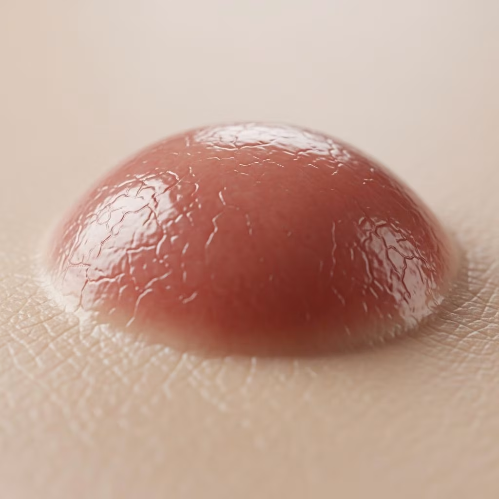
A Spitz naevus is a benign skin tumour. However, it may resemble a melanoma clinically and microscopically, so Spitz naevi are often excised as a precaution.
What is a Spitz naevus?
A Spitz naevus (also called Spitz nevus) is a type of melanocytic naevus, that is, it is a mole composed of melanocytes — these are cells that normally produce pigment, melanin, and are responsible for skin colour. The melanocytes in Spitz naevus may be inactive. Thus the lesion may be pink rather than brown.
The genetic pattern of the DNA in Spitz naevus is characteristic with kinase fusions being prominent.
It is not known why Spitz naevi occur.
Who gets Spitz naevi?
Spitz naevi are seen most often in children; 70% of cases diagnosed during the first 20 years of life. They may also arise in adults. They are most frequently found in fair-skinned individuals (skin phototypes I and II) but can also affect those with dark skin.
Types of Spitz naevus
Spitz naevus is classified as:
- Classic Spitz naevus
- Pigmented Spitz naevus
- Spindle cell tumour of Reed
There are clinical features in common for all three types of Spitz naevus.
The atypical Spitz naevus or spitzoid naevus is so called because its features differ from those seen in the majority of Spitz naevi.
The rare entity, agminated Spitz naevus, presents as multiple Spitz naevi in a segmental presentation. It is an example of cutaneous mosaicism and is due to a genetic mutation.
Clinical features
A Spitz naevus is often suspected clinically by its characteristic dome-shaped appearance and rapid growth over a few weeks to months.
- Usually 2–6 mm in diameter, but atypical examples may be larger
- Solitary lesion
- Symmetrical dome shaped or smooth surface
- Light to dark pink, reddish-brown to black
- Sometimes scaly or crusted surface
- Rapid growth over weeks to months
- Legs and face most common sites
In older children and adults, the diagnosis of Spitz naevus is usually confirmed by an excisional skin biopsy.
Classic Spitz naevus
Classic Spitz naevi are often dome-shaped and reddish-brown to pinkish, but they may also be colourless. They typically appear on the face, neck, or legs, grow quickly, and may itch, bleed, or ooze.
Pigmented Spitz naevus
Pigmented Spitz naevi may be light to dark brown or black. They are frequently found on the extremities and can mimic acral lentiginous melanoma in rare cases.
Spindle cell tumour of Reed
This variant features fusiform cells and can co-exist with other forms. It presents with similar clinical features but distinct histology.
Atypical Spitz naevus
Atypical features include:
- Larger size (>1 cm)
- Irregular borders
- Variable colour
- Scaly or ulcerated surface
- Often on the back
Agminated Spitz naevus
Multiple lesions confined to a segmental area due to mosaicism.
Diagnosis
The pathology of Spitz naevus usually shows a symmetrical compound naevus composed of nests of characteristic epithelioid cells in classic and pigmented Spitz, and fusiform cells in spindle cell tumour of Reed. Both forms can co-exist.
Dermoscopy reveals patterns such as starburst (peripheral pseudopods) or globular (large globules).
Differential diagnosis includes melanoma, especially atypical cases. Biopsy is essential in adults.
What is the treatment for Spitz naevus?
In children, small classic Spitz naevi may be left alone if the clinical diagnosis is certain. However, excision is often recommended due to diagnostic uncertainty.
In adults, excisional biopsy is standard to rule out melanoma.
Atypical Spitz tumours may require wider excision and sentinel lymph node biopsy in select cases.
Complications
Spitz naevi are benign but can cause anxiety due to melanoma mimicry. Rare malignant transformation reported in atypical variants.
Prevention
No known prevention. Regular skin checks recommended, especially in fair-skinned individuals.
| Feature | Spitz Naevus | Melanoma |
|---|---|---|
| Age | Mostly <20 years | Any age, peaks later |
| Growth | Rapid then stable | Progressive |
| Symmetry | Symmetrical | Asymmetrical |
| Histology | Characteristic cells, maturation | Atypical mitosis, invasion |
Frequently asked questions
Is Spitz naevus cancerous?
No, Spitz naevus is benign, though it mimics melanoma and is often excised.
Does Spitz naevus itch or bleed?
Yes, it may itch, bleed, or ooze, especially during rapid growth phase.
Can Spitz naevus appear in adults?
Yes, though less common than in children. Biopsy recommended.
How is Spitz naevus diagnosed?
By clinical exam, dermoscopy, and excisional biopsy with histopathology.
Do Spitz naevi disappear?
Some may flatten or regress over years, but excision is common.
This article provides an in-depth exploration of Spitz naevus, expanding on clinical variants, diagnostic challenges, and management strategies. While primarily affecting children, awareness is crucial for all ages due to its melanoma-like appearance. Early recognition through ABCDE criteria (Asymmetry, Border irregularity, Color variation, Diameter >6mm, Evolving) aids differentiation, though biopsy remains gold standard. Genetic insights into kinase fusions offer future diagnostic promise. Regular dermatological surveillance is advised for at-risk populations.
References
- Spitz naevus (Spitz nevus) – DermNet — DermNet NZ. 2023. https://dermnetnz.org/topics/spitz-naevus
- Spitz Nevus: Identification, Treatment, Pictures, and More — Healthline. 2023-05-12. https://www.healthline.com/health/spitz-nevus
- Spitz naevus — Cancer Research UK. 2024. https://www.cancerresearchuk.org/about-cancer/other-conditions/spitz-naevus
- Spitz nevus: Pictures, diagnosis, and treatment — Medical News Today. 2023-09-15. https://www.medicalnewstoday.com/articles/320805
- Acral Pigmented Spitz Nevus That Clinically Mimicked Acral… — NIH PMC. 2011-06-27. https://pmc.ncbi.nlm.nih.gov/articles/PMC3130876/



