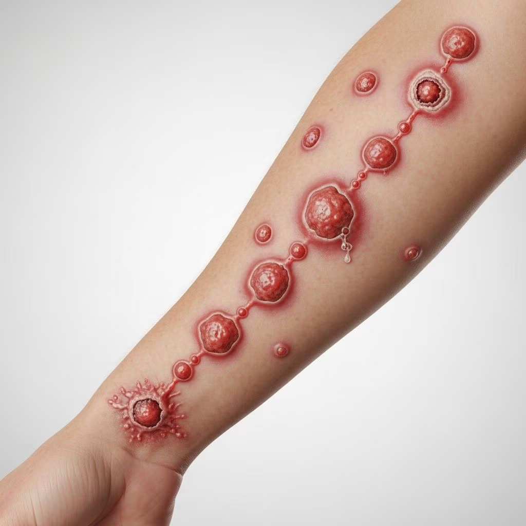
Sporotrichoid lymphocutaneous infection, also known as nodular lymphangitis, is a distinctive dermatological syndrome characterized by a linear arrangement of cutaneous nodules or ulcers ascending along superficial lymphatic channels from a primary lesion. This pattern typically follows minor skin trauma or inoculation, most commonly affecting the extremities.
What is Sporotrichoid Lymphocutaneous Infection?
The term “sporotrichoid” derives from sporotrichosis, the prototype infection caused by the dimorphic fungus Sporothrix schenckii, which classically produces this lymphatic spread pattern. The syndrome involves granulomatous inflammation progressing linearly along lymphatics, often triggered by traumatic inoculation of pathogens from soil, plants, water, or animals.
Lesions begin as a primary nodule at the inoculation site, followed by secondary nodules spaced 2-3 cm apart along draining lymphatics. Each nodule is firm, erythematous, and may suppurate, ulcerate, or form abscesses. Regional lymphadenopathy is common, but systemic symptoms are typically absent unless disseminated.
Who Gets Sporotrichoid Lymphocutaneous Infection?
This infection affects individuals with outdoor occupations or hobbies involving soil, plants, or water exposure, such as gardeners, farmers, fishermen, and aquarium enthusiasts. It occurs worldwide but shows regional variations: Sporothrix spp. predominate globally, while nontuberculous mycobacteria (NTM) like Mycobacterium marinum are common in Asia, and dematiaceous fungi prevail in parts of Thailand.
Immunocompromised patients (e.g., HIV/AIDS, diabetes, corticosteroids) are at higher risk for atypical or disseminated forms, though most cases occur in immunocompetent hosts. Children and adults are equally affected, with no strong gender predilection.
Causes
Sporotrichoid pattern arises from diverse pathogens capable of lymphatic spread after cutaneous inoculation. Major categories include:
- Fungi: Sporothrix schenckii (classic, associated with rose thorns, plant material); dematiaceous fungi (Exophiala spp., Wangiella spp., Fonsecaea spp.) increasingly reported in Asia.
- Mycobacteria: M. marinum (fish tanks, water); M. chelonae, M. fortuitum, M. haemophilum (rapid growers).
- Bacteria: Nocardia brasiliensis, N. asteroides (soil); rarely Staphylococcus aureus, Streptococcus pyogenes, Francisella tularensis (tularemia).
- Parasites: Leishmania spp. (endemic areas).
- Rare/Noninfectious: Metastatic carcinoma, sarcoidosis.
In a 10-year Thai study, dematiaceous fungi were identified in 58% of culture-positive cases, surpassing Sporothrix (4%).
Clinical Features
The incubation period varies: days for bacteria (e.g., tularemia), 1-2 weeks for fungi/mycobacteria.
- Primary lesion: Painless papule/nodule at trauma site (e.g., finger, leg), evolving to pustule/ulcer (1-2 cm).
- Lymphatic spread: Chain of 3-10 nodules (0.5-2 cm) along lymphatics, spaced linearly; may fluctuate, ulcerate, or fistulize.
- Skin changes: Erythema, tenderness; no fever/lymphadenitis in fungal/mycobacterial cases, unlike tularemia.
- Sites: Upper > lower limbs; rarely trunk/face.
Differential by history:
| Exposure | Likely Pathogen |
|---|---|
| Plants/thorns | Sporothrix, dematiaceous fungi |
| Aquariums/water | M. marinum |
| Soil/trauma | Nocardia, rapid-grower mycobacteria |
| Animals/bites | Tularemia, sporotrichosis (cats) |
Diagnosis
Diagnosis combines history, exam, microbiology, and histopathology; empirical therapy risks missing treatable causes.
- Investigations:
- Skin biopsy: Punch/full-thickness for histopathology (H&E, special stains: PAS, GMS for fungi; Fite/Ziehl-Neelsen for mycobacteria; Gram/Wade-Fite for bacteria/Nocardia).
- Culture: Essential; fungal (Sabouraud), mycobacterial (Lowenstein-Jensen), bacterial blood agar. Yield ~45%.
- Molecular: PCR for mycobacteria/Leishmania if available.
- Histopathology: Granulomas (suppurative/granulomatous); organisms visible in <50% (asteroid bodies in sporotrichosis rare).
Skin biopsy is crucial for suspicious cases, guiding therapy.
Treatment
Treatment targets identified pathogen; empirical itraconazole for suspected fungal (esp. pet/plant exposure).
| Pathogen | First-line Therapy | Duration |
|---|---|---|
| Sporothrix schenckii | Itraconazole 200 mg/day | 3-6 months |
| Dematiaceous fungi | Itraconazole 200-400 mg/day | 6-12 months |
| M. marinum | Rifampin + ethambutol or clarithromycin | 3-6 months |
| Rapid-grower mycobacteria | Clarithromycin + amikacin/ethambutol | 4-6 months |
| Nocardia | Trimethoprim-sulfamethoxazole | 6-12 weeks |
Surgical excision/drainage adjunctive; monitor response clinically/culture. Poor prognostic factors: immunosuppression, delayed diagnosis.
What is the Outcome?
With appropriate therapy, cure rates exceed 90%; relapse rare if full course completed. Untreated, chronic ulceration/scarring; dissemination in immunocompromised (osteoarticular, pulmonary). In Thailand series, most responded to antifungals.
Prevention
- Wear gloves/protective clothing for gardening, handling fish/plants.
- Clean wounds promptly; avoid non-chlorinated water if cut.
- Avoid cat scratches (Sporothrix zymosis emerging).
Frequently Asked Questions
Is sporotrichoid infection contagious?
No, it spreads via inoculation/trauma, not person-to-person.
How long does it take to develop after injury?
Days (bacteria) to weeks (fungi/mycobacteria).
Can it be cured without antibiotics/antifungals?
Rarely; most require prolonged systemic therapy.
What if biopsy is negative?
Treat empirically based on exposure; repeat biopsy or monitor.
Is it common in travelers?
Yes, from endemic areas (tropics, Asia).
References
- A 10-Year Retrospective Study of Sporotrichoid Lymphocutaneous Infection in Southern Thailand — Phoompoung P, et al. 2023-04-15. https://pmc.ncbi.nlm.nih.gov/articles/PMC10076999/
- Sporotrichoid Lymphocutaneous Infection — DermNet NZ. 2023. https://dermnetnz.org/topics/sporotrichoid-lymphocutaneous-infection
- Sporotrichoid Lymphocutaneous Spread of Metastatic Cutaneous Squamous Cell Carcinoma — UNTHSC Scholarly Repository. 2020. https://unthsc-ir.tdl.org/items/2b330056-adf3-4271-9592-55b06087279f
- Sporotrichoid Lymphocutaneous Infections: A Review — American Family Physician (AAFP). 2001-01-15. https://www.aafp.org/pubs/afp/issues/2001/0115/p326.html
- Nodular Lymphangitis (Sporotrichoid Lymphocutaneous Infections): Review — PMC. 2018-06-29. https://pmc.ncbi.nlm.nih.gov/articles/PMC6023502/
- Sporotrichosis — Merck Manuals Professional Edition. 2024. https://www.merckmanuals.com/professional/infectious-diseases/fungal-infections/sporotrichosis
- Sporotrichosis Fact Sheet — World Health Organization (WHO). 2023-11-10. https://www.who.int/news-room/fact-sheets/detail/sporotrichosis