
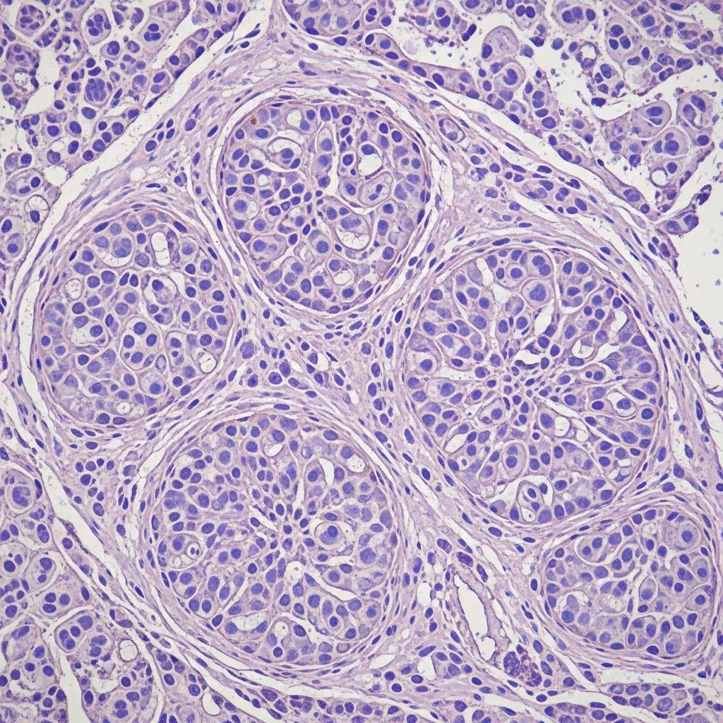
Squamous cell carcinoma (SCC) represents a common keratinocytic skin cancer primarily linked to ultraviolet radiation exposure from sunlight. Typical SCC features nests of squamous epithelial cells originating from the epidermis and invading the dermis. These malignant cells exhibit large size, abundant eosinophilic cytoplasm, and prominent vesicular nuclei, often with variable keratinisation forming keratin pearls.
Introduction
Cutaneous squamous cell carcinoma is the second most prevalent skin malignancy in the United States, arising from keratinocytes in the epidermis. It develops due to DNA mutations, notably in the tp53 tumor suppressor gene, common in both SCC and its precursor actinic keratosis. Immunosuppression significantly elevates risk, with solid organ transplant patients facing 65- to 250-fold higher incidence compared to the general population.
SCC typically manifests on sun-exposed areas like the face, scalp, ears, and hands but can occur anywhere, including less exposed sites in darker skin types. While often non-life-threatening, untreated cases can invade locally or metastasise, particularly high-risk variants.
Clinical features
SCC presents as firm, red nodules or plaques, often with scaling, ulceration, or crusting. Common sites include sun-damaged skin on the head, neck, dorsum of hands, and lips. Lesions may grow rapidly or persist as non-healing sores. In immunosuppressed individuals or those with chronic wounds, SCC can arise in scars or burns (Marjolin ulcer).
- Rough, scaly patches resembling warts or actinic keratosis.
- Open sores with raised, rolled borders.
- Persistent, tender nodules that bleed easily.
- Intraoral or genital involvement in high-risk cases.
Pathogenesis
UV radiation induces DNA damage in squamous cells, leading to uncontrolled proliferation. Key mutations affect tp53, disrupting cell cycle regulation. Chronic inflammation, HPV infection, and immunosuppression further promote oncogenesis. In fair-skinned individuals, low melanin exacerbates UV damage.
Precursors include actinic keratosis (AK) and Bowen disease (SCC in situ), where atypia is confined to the epidermis. Invasion beyond the basal layer defines invasive SCC.
Cytological features
| Feature | Description |
|---|---|
| Cell size | Large atypical keratinocytes |
| Cytoplasm | Abundant eosinophilic, often keratinising |
| Nucleus | Vesicular with prominent nucleoli |
| Border | Irregular, hyperchromatic |
| Keratinisation | Individual cell or pearl formation |
Malignant squamous cells display marked atypia: increased nuclear-to-cytoplasmic ratio, pleomorphism, and mitotic figures. Keratin production yields eosinophilic pearls, a hallmark feature distinguishing SCC from other epithelia-derived tumours.
Histological variants
SCC exhibits diverse histological patterns influencing prognosis. Variants range from low-risk (e.g., AK-associated) to high-risk (e.g., desmoplastic).
Conventional SCC
Lobular architecture with irregular nests penetrating the dermis. Tumour islands show peripheral palisading and central keratinisation. Stroma may be desmoplastic or inflamed.
Acantholytic SCC
Characterised by suprabasal clefting and acantholysis, mimicking pseudoglandular spaces. Less cohesive cells with gland-like structures; higher recurrence risk.
Adenosquamous carcinoma / mucoepidermoid carcinoma
Rare glandular differentiation, predominantly on head and neck. Mucin production within squamous nests; aggressive behaviour.
Desmoplastic SCC
Infiltrative growth in dense, fibrous stroma without clear nests. High recurrence and metastasis rates due to subtle invasion.
Infiltrative SCC
Small nests, strands, or single cells infiltrating fibrous/mucinous dermis. Subtle borders challenge margin assessment.
Metaplastic SCC (carcinosarcoma)
Biphasic: malignant epithelial (SCC) and mesenchymal (sarcoma-like) components. Exceedingly rare, aggressive.
Pigmented SCC
Melanin in tumour cells, macrophages, or melanocytes. Distinguished from squamomelanocytic tumours.
Spindle cell SCC
Pleomorphic spindle cells with minimal keratinisation. Often radiation-associated; immunostains (p63, cytokeratins) confirm epithelial origin.
Other variants
- Clear cell SCC: Glycogen-rich clear cytoplasm.
- Signet ring SCC: Intracytoplasmic mucin displacing nucleus.
- Lymphoepithelioma-like: Lymphoid stroma; HPV-related.
High-risk features
Prognostic indicators include:
- Depth >2mm or Clark level IV/V.
- Perineural invasion (especially >0.1mm calibre nerves).
- Lymphovascular invasion.
- Poor differentiation.
- Immunosuppression or tumour diameter >2cm.
Differential diagnosis
| Entity | Key Distinguishing Features |
|---|---|
| Keratoacanthoma | Symmetrical crateriform architecture, overhanging lips |
| Basal cell carcinoma | Basaloid cells, peripheral palisading, mucinous stroma |
| Merkel cell carcinoma | Small round cells, CK20+, neuroendocrine markers |
| Melanoma | S100+, melanocytic markers; junctional activity |
| Adnexal carcinoma | Ductal/lobular patterns, decapitation secretion |
Treatment and prognosis
Surgical excision is primary, with Mohs micrographic surgery preferred for high-risk sites (lips, ears) or recurrent lesions. Factors include size >2cm, high-risk histology, and immunosuppression. Radiation or systemic therapy for advanced/metastatic disease.
Low-risk SCC: <2% metastasis. High-risk: >10%, especially de novo, post-radiation, or desmoplastic types. Early detection yields excellent cure rates.
Frequently asked questions
What causes squamous cell carcinoma?
Primarily UV exposure; also immunosuppression, chronic wounds, HPV, fair skin.
Is SCC life-threatening?
Rarely, if treated early; high-risk features increase metastasis risk.
How is SCC diagnosed?
Biopsy with histopathology; Mohs for margins in select cases.
What are common treatments?
Excision, Mohs surgery, curettage, radiation.
Can SCC recur?
Yes, especially infiltrative or desmoplastic variants; regular follow-up essential.
References
- Squamous cell carcinoma of the skin – Symptoms and causes — Mayo Clinic. 2023-10-15. https://www.mayoclinic.org/diseases-conditions/squamous-cell-carcinoma/symptoms-causes/syc-20352480
- Squamous cell carcinoma pathology — DermNet NZ. 2024-01-01. https://dermnetnz.org/topics/squamous-cell-carcinoma-pathology
- Squamous Cell Carcinoma — Cockerell Dermatopathology. 2023-05-20. https://www.dermpath.com/squamous-cell-carcinoma
- Cutaneous Squamous Cell Carcinoma — StatPearls, NCBI Bookshelf. 2024-07-25. https://www.ncbi.nlm.nih.gov/books/NBK441939/
- Cutaneous squamous cell carcinoma: a comprehensive clinicopathologic classification — Journal of Cutaneous Pathology, Wiley. 2006-11-01. https://onlinelibrary.wiley.com/doi/10.1111/j.0303-6987.2006.00516.x



