
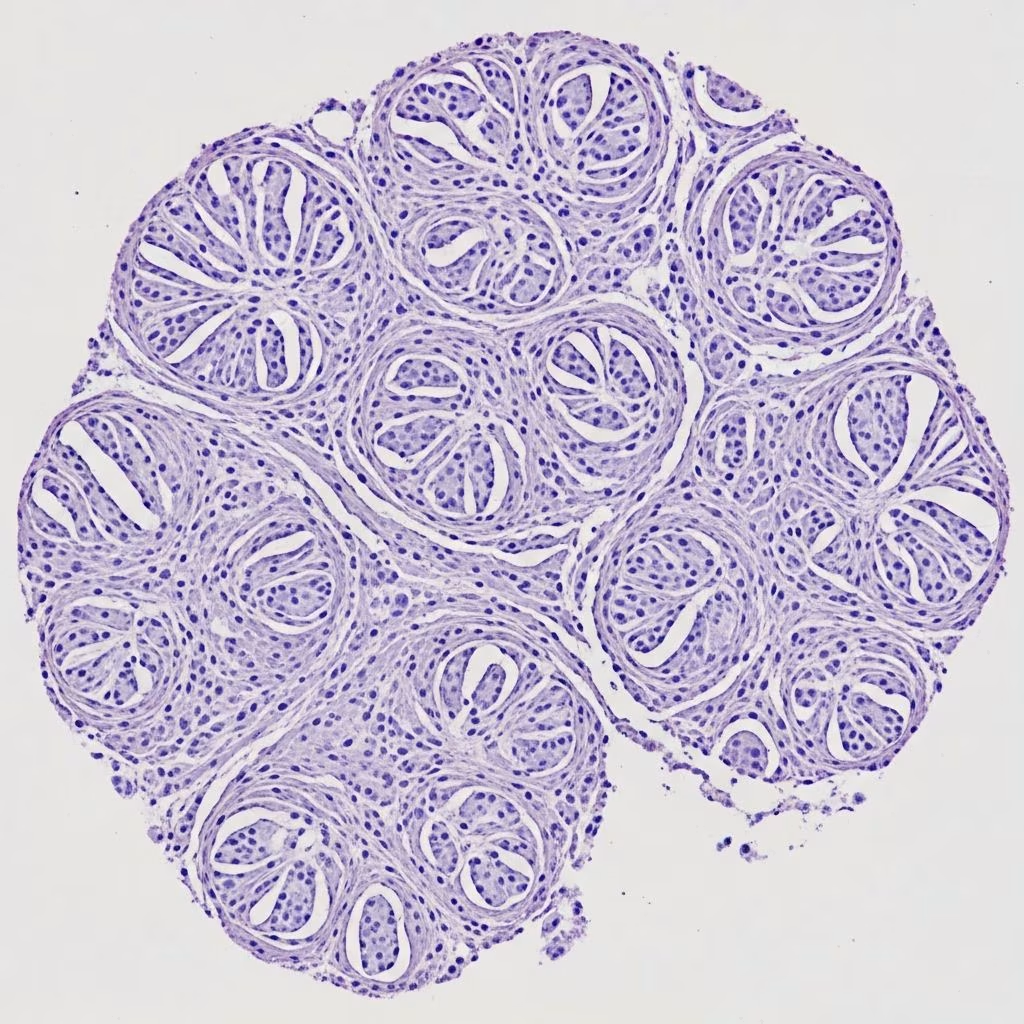
Superficial acral fibromyxoma (SAF), also known as digital fibromyxoma, is a rare, benign soft tissue neoplasm with a strong predilection for the acral sites, particularly the digits of hands and feet. First described in 2001, this tumour typically presents as a slow-growing, solitary nodule in the periungual or subungual regions, often mimicking more common conditions like warts or onychomycosis. SAF is characterized histologically by spindled to stellate fibroblasts within a myxoid or collagenous stroma, with prominent vascularity and immunoreactivity for CD34. Although benign, incomplete excision can lead to local recurrence in up to 22% of cases. This article details the clinical, histological, and immunohistochemical features essential for accurate diagnosis and management.
Introduction
Superficial acral fibromyxoma represents a distinct entity among fibroblastic/myofibroblastic tumours, distinguished by its location and morphological features. It arises in the dermis and superficial subcutis of acral skin, with over 300 cases reported worldwide since its initial description by Fetsch et al. in 2001. The tumour shows a slight male predominance (2:1 ratio) and affects a wide age range (4–86 years), though most commonly middle-aged adults. Clinically, SAF manifests as a firm, painless nodule, often longstanding and unnoticed until trauma or growth prompts evaluation. Its rarity and nonspecific appearance contribute to frequent misdiagnosis, underscoring the need for histopathological confirmation.
Terminology
- Superficial acral fibromyxoma (preferred term)
- Digital fibromyxoma
- Acral fibromyxoma
These synonyms reflect its characteristic location on digits and acral surfaces.
Clinical features
Epidemiology
- Incidence: Rare; approximately 326 cases reported globally.
- Age: Wide range (4–86 years); peak >40 years.
- Sex: Male predominance (M:F ≈ 2:1).
Sites
- Almost exclusively acral: hands (fingers > toes) and feet.
- Preferred locations: Periungual (50%) and subungual regions; great toe and index finger common.
- Rarely palm, heel, or ankle.
Clinical presentation
- Solitary, slow-growing nodule (0.6–5 cm; median 1.5 cm).
- Firm, flesh-coloured to pink; often asymptomatic but pain in 40–50%.
- Nail involvement: onycholysis, dystrophy, hyperkeratosis, or lunula deformity.
- May erode bone (scalloping on imaging).
Dermoscopy may show whitish structureless areas, linear vessels, or microhemorrhages in subungual cases.
Histopathology
Macroscopic
- Well-circumscribed but unencapsulated; soft-firm, gelatinous white-gray.
- Size typically <2 cm; no necrosis or haemorrhage.
Microscopic
SAF is a dermal-based, multilobulated tumour extending superficially into subcutis. Key features include:
- Cells: Bland spindled/stellate fibroblasts; minimal nuclear atypia; rare mitoses (<1/10 HPF).
- Architecture: Loose fascicles, storiform or haphazard arrays.
- Stroma: Myxoid to collagenous; alternating myxoid/fibrotic areas.
- Vascularity: Prominent thin-walled vessels.
- Other: Mast cells frequent; occasional multinucleated cells; rare bone erosion.
Low-power view shows a multinodular dermal proliferation with extension into subcutis. Medium-power reveals fascicular/storiform spindle cells in myxocollagenous stroma. High-power demonstrates bland nuclei, fine chromatin, and small nucleoli amid delicate vessels.
Histopathology images
- Scanning power: Multilobular dermal tumour with subcutaneous extension.
- Low power: Fascicles of spindle cells in myxoid stroma with vascularity.
- Medium power: Storiform pattern with collagen trapping.
- High power: Stellate cells, mast cells, and thin vessels.
- Prominent collagen: Fibrous variant with thick bundles.
Immunohistochemistry
| Antibody | Reactivity | Pattern |
|---|---|---|
| CD34 | Positive (most cases) | Diffuse to focal; cytoplasmic |
| CD99 | Positive | Membranous |
| Vimentin | Positive | Diffuse |
| CD10 | Often positive | Cytoplasmic |
| EMA | Focal (rare) | – |
| S100 | Negative | – |
| Desmin | Negative | – |
| Keratins | Negative | – |
| Claudin-1 | Negative | – |
CD34 positivity is characteristic but not diagnostic alone; combination with clinical/histological features required.
Genetics
- Non-specific changes; no recurrent mutations identified.
- Possible STAT6 negativity distinguishes from DFSP.
Differential diagnosis
| Diagnosis | Key Distinguishing Features |
|---|---|
| Dermatofibroma | Epidermal hyperplasia; epidermal-dermal interface; more storiform; factor XIIIa+ |
| Perineurioma | Slender processes; prominent vessels; EMA+, claudin-1+ |
| Digital fibrokeratoma | Collagen core with vessels; epidermal collarettes |
| DFSP | Infiltrative; storiform; CD34+; t(17;22) |
| Myxoid neurofibroma | S100+; wavy nuclei; inherited association |
| Onychomatricoma | Matrix origin; filamentous digits |
| Epidermal inclusion cyst | Keratin-filled; epithelial lining |
Management
- Treatment: Complete surgical excision with clear margins.
- Prognosis: Excellent; low recurrence if fully excised (up to 22% with incomplete removal).
- Avoid overtreatment (e.g., amputation); MRI for bone involvement.
Currently used pathology tests
- Histology (H&E)
- CD34, CD99 immunohistochemistry
- Consider S100, EMA, STAT6 to exclude mimics
Main DDx
- Perineurioma
- Dermatofibroma
- DFSP
Download slides
(PDF slides available for educational use)
Frequently asked questions
Q: Is superficial acral fibromyxoma malignant?
No, SAF is entirely benign with no metastatic potential.
Q: What is the most common site for SAF?
Periungual and subungual regions of fingers and toes, especially great toe.
Q: Is CD34 specific for SAF?
No; CD34 is supportive but requires clinicohistological correlation.
Q: Does SAF recur frequently?
Rare if completely excised; up to 22% with incomplete removal.
Q: Can SAF involve bone?
Yes, bone scalloping seen on imaging in some cases.
References
- Superficial acral fibromyxoma — JL Sawaya et al. International Journal of Dermatology. 2015-03-23. https://pubmed.ncbi.nlm.nih.gov/25772615/
- Superficial acral fibromyxoma — Wikipedia (sourced from primary refs). N/A. https://en.wikipedia.org/wiki/Superficial_acral_fibromyxoma
- Superficial acral fibromyxoma: insights from case analysis of 326 reported cases — HL Tan et al. PMC. 2024. https://pmc.ncbi.nlm.nih.gov/articles/PMC10873247/
- Superficial Acral Fibromyxoma — Basic Medical Key. N/A. https://basicmedicalkey.com/superficial-acral-fibromyxoma-3/
- Superficial acral fibromyxoma with bone involvement — Anais de Dermatologia. 2024. https://www.anaisdedermatologia.org.br/en-superficial-acral-fibromyxoma-with-bone-articulo-S0365059624002629
- Superficial acral fibromyxoma: Clinical, dermoscopic and reflectance confocal microscopy features — M Starace et al. Journal of the European Academy of Dermatology and Venereology. 2023. https://onlinelibrary.wiley.com/doi/10.1111/jdv.19101



