
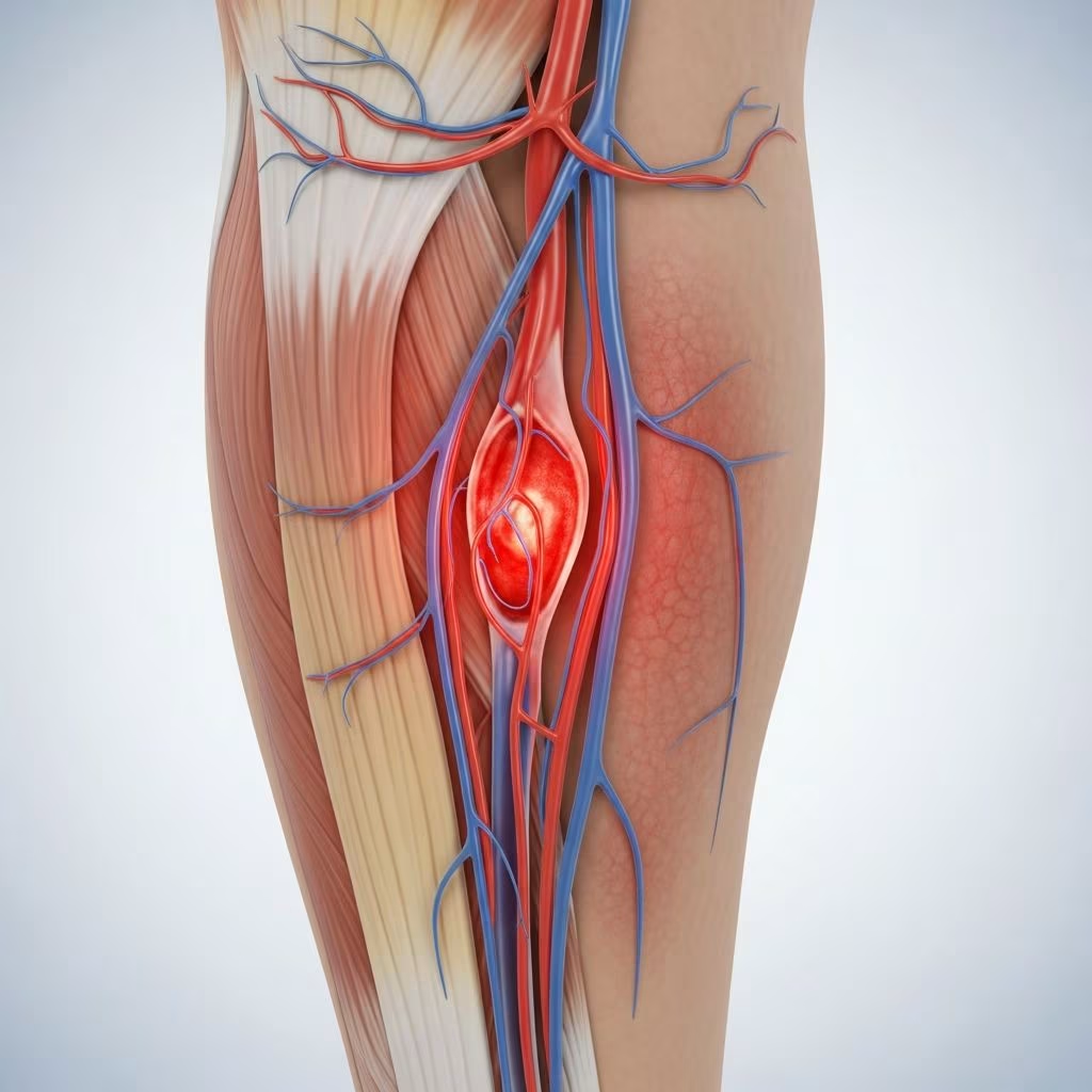
Superficial thrombophlebitis involves inflammation and clotting in veins close to the skin’s surface, most often in the legs. This condition typically resolves without complications but requires monitoring to prevent extension into deeper veins.
Defining Superficial Thrombophlebitis
Superficial thrombophlebitis, also called superficial vein thrombosis (SVT), occurs when a blood clot forms in a superficial vein alongside inflammation. These veins lie just under the skin, unlike deeper ones surrounded by muscle. It primarily affects the lower extremities, with 60-80% of cases involving the great saphenous vein along the inner leg.
The condition arises from a combination of clot formation (thrombosis) and vein wall irritation (phlebitis). While generally benign, involvement near deep vein junctions can signal higher risks, such as progression to deep vein thrombosis (DVT).
Common Locations and Patterns
- Great saphenous vein: Most frequent site, running from groin to ankle.
- Small saphenous vein: Affects 10-20% of cases, along the outer calf.
- Arm veins: Less common, often from IV insertions.
- Recurrent episodes: Prior occurrences raise future risk.
Root Causes and Risk Factors
Several factors contribute to superficial thrombophlebitis development. Varicose veins top the list, present in 75-88% of cases, as weakened valves cause blood pooling and stasis.
Other triggers include:
- Vein trauma from injections, cannulas, or injury.
- Hypercoagulable states like thrombophilia or estrogen use.
- Prolonged immobility, obesity, or recent surgery.
- Autoimmune issues such as Behçet’s disease or infections.
- Dehydration or turbulent flow in diseased veins.
| Risk Factor | Description | Prevalence |
|---|---|---|
| Varicose Veins | Enlarged, twisted veins with poor flow | 75-88% |
| IV/Injection Trauma | Direct vein irritation | Common in hospitalized patients |
| Immobility | Long travel or bed rest | Increases stasis risk |
| Hypercoagulability | Genetic or acquired clotting tendency | Associated with recurrence |
Pathophysiology begins with microscopic clots from stasis, injury, or coagulopathy, per Virchow’s triad. Endothelial damage triggers platelet adhesion and thrombin activation, forming macroscopic thrombi.
Recognizing Symptoms Early
Patients often notice a firm, tender cord-like vein under red, warm skin. Surrounding swelling, itching, or mild fever may accompany it.
- Localized pain intensifies with touch or movement.
- Red streak traces the vein path.
- Low-grade inflammation without systemic signs.
- Distinguish from DVT: No major leg swelling unless extension occurs.
Symptoms peak within days and fade over 1-2 weeks if uncomplicated.
How It’s Diagnosed
Diagnosis relies on clinical exam and ultrasound to confirm clot location and rule out DVT. Palpation reveals the indurated cord.
Duplex ultrasound is gold standard, assessing clot extent and deep vein involvement. It’s non-invasive and highly accurate.
- History review: Recent trauma, varicose veins, or clotting disorders.
- Physical: Inspect for erythema, edema, and cord.
- Imaging if extensive or near saphenofemoral junction.
Extensive SVT or no clear cause may prompt thrombophilia screening, though evidence is inconclusive.
Treatment Approaches
Management focuses on symptom relief and preventing propagation. Most cases self-resolve in weeks.
Conservative Measures
- Elevate affected limb to reduce swelling.
- Apply warm compresses for comfort.
- Compression stockings improve flow.
Medications
Nonsteroidal anti-inflammatory drugs (NSAIDs) like ibuprofen ease pain and inflammation. Topical NSAIDs offer localized relief.
Anticoagulants (fondaparinux, low-molecular-weight heparin) for high-risk cases near deep veins or extensive clots.
Interventional Options
Saphenous ligation or vein ablation if recurrent or linked to varicosities.
| Treatment | Use Case | Duration |
|---|---|---|
| NSAIDs | Pain/inflammation | 1-2 weeks |
| Compression | All cases | Ongoing |
| Anticoagulants | High-risk SVT | 45 days |
| Surgery | Recurrent/near junction | As needed |
When to Worry: Complications
Rarely, SVT extends to deep veins (5.4% risk for DVT/PE). Monitor if clot nears saphenofemoral junction.
- Signs of extension: Increasing swelling, severe pain.
- Migratory pattern: Suggests underlying malignancy.
- Chronic cases: May lead to post-thrombotic changes.
Prevention Strategies
Address modifiable risks:
- Manage varicose veins via lifestyle or treatment.
- Stay active; avoid prolonged sitting.
- Hydrate during travel; use compression socks.
- Minimize IV trauma in medical settings.
For high-risk individuals, discuss prophylactic measures with providers.
Superficial vs. Deep Vein Issues
| Aspect | Superficial Thrombophlebitis | Deep Vein Thrombosis (DVT) |
|---|---|---|
| Location | Surface veins | Deep veins |
| Risk Level | Low, self-limiting | High (embolism risk) |
| Symptoms | Local redness, cord | Leg swelling, heaviness |
| Treatment | NSAIDs, compression | Anticoagulants urgent |
Living with Vein Health Concerns
Superficial thrombophlebitis signals potential venous insufficiency. Long-term, strengthen legs with exercise, maintain weight, and elevate legs. Regular check-ups benefit those with varicosities.
Women on hormones or post-surgery patients should watch for early signs.
Frequently Asked Questions (FAQs)
Is superficial thrombophlebitis dangerous?
Usually not; it resolves quickly. But seek care if near deep veins.
How long does it last?
1-2 weeks with treatment.
Can it recur?
Yes, especially with varicose veins.
Do I need blood thinners?
Only for extensive or high-risk cases.
Can exercise help?
Yes, promotes circulation; avoid during acute phase.
References
- Superficial thrombophlebitis — Dr Johan Blignaut, Vein Surgery. Accessed 2026. https://www.veinsurgery.co.za/superficial-thrombophlebitis.html
- Phlebitis (superficial thrombophlebitis) — NHS. 2023-02-15. https://www.nhs.uk/conditions/phlebitis/
- Superficial Thrombophlebitis — StatPearls, NCBI Bookshelf, NIH. 2023-08-08. https://www.ncbi.nlm.nih.gov/books/NBK556017/
- What You Should Know About Superficial Thrombophlebitis — Vanishing Veins. Accessed 2026. https://vanishingveins.net/surface-vein-clots-what-you-should-know-about-superficial-thrombophlebitis/
- Superficial Thrombophlebitis: Symptoms & Causes — Cleveland Clinic. 2023-11-01. https://my.clevelandclinic.org/health/diseases/17523-superficial-thrombophlebitis
- How superficial thrombophlebitis develops — YouTube (health video). Accessed 2026. https://www.youtube.com/watch?v=1N_bxIzFGOI
- Superficial Thrombophlebitis — University of Michigan Health. Accessed 2026. https://www.uofmhealth.org/our-care/specialties-services/superficial-thrombophlebitis



