
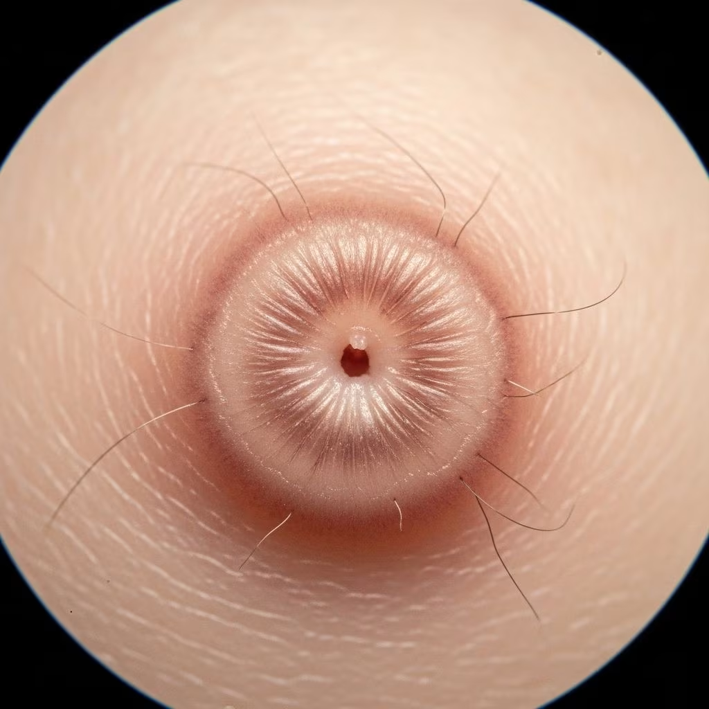
A supernumerary nipple, also known as an accessory or ectopic nipple, represents a minor congenital malformation of mammary tissue, resulting in an extra nipple with potential associated glandular structures. These anomalies occur due to incomplete regression of embryonic milk lines and are present from birth, often mimicking benign skin lesions like naevi.
Introduction
Supernumerary nipples arise from the failure of mammary ridges to regress during embryonic development. These ridges, or milk lines, extend bilaterally from the axilla to the groin. Normally, only the thoracic pair persists to form the primary nipple-areola complex, while others involute. Persistence leads to extra nipples, reported in up to 6% of the population, though many go unnoticed due to their small size.
These lesions are typically asymptomatic but warrant recognition to rule out associated malformations or rare malignancies. Dermoscopy plays a crucial role in non-invasive diagnosis, revealing characteristic patterns that distinguish them from melanocytic or fibrotic lesions.
Clinical Features
Supernumerary nipples manifest at birth as small, pink or brown macules or papules along the milk lines, most commonly below the normal breast in the inframammary region, axilla, or abdomen. They measure 1-2 cm, smaller than typical nipples, with a central elevation or dimple resembling a nipple tip. Puberty may induce pigmentation, hair growth, or glandular enlargement, simulating Montgomery tubercles or naevi.
- Location: Primarily along milk lines (axilla to groin); 5% ectopic on neck, back, thigh, or vulva.
- Appearance: Firm plaque with elevated central nipple-like projection; may be solitary or multiple, unilateral or bilateral.
- Evolution: Stable but can hypertrophy at puberty or lactation; rarely lactate.
- Associations: Familial in 6% (autosomal dominant); linked to syndromes like Simpson-Golabi-Behmel or urinary tract anomalies (disputed).
In adults, they may present as pigmented lesions prompting biopsy if unrecognized, as in a case of a 40-year-old woman with breast cancer history showing a 2 cm brown plaque under the breast.
Dermoscopic Features
Dermoscopy of supernumerary nipples characteristically shows pigment network-like structures peripherally with central scar-like or cleft features, aiding differentiation from melanoma or dermatofibroma. These patterns reflect underlying epidermal and dermal architecture.
| Dermoscopic Feature | Definition | Frequency/Notes |
|---|---|---|
| Peripheral network-like structures | Brownish interconnected thin lines over tan background at lesion periphery | Most common; seen in all cases with central dimpling |
| Central network-like structures | Similar network centrally | Often with white cobblestones |
| Central white scar-like area | Sharply circumscribed round/oval white patch centrally | Classic; mimics dermatofibroma |
| Cleft-like appearance | Small rim dividing tip into symmetrical parts | 42% of cases |
| White cobblestone-like structures | Aggregated angulated whitish globules | Associated with central network |
| Central round dimpling with plug | Round dimple with darker central plug | 31%; alternative to cleft |
| Fisheye-like structures | Dark brown/black round dots mimicking comedos | Newly described |
Studies confirm pigment network in 100% of cases, with peripheral type predominant. Combination of peripheral network and central scar-like area is highly suggestive. Variations include atrophic dermatofibroma mimicry with central white patch.
Differential Diagnoses
Supernumerary nipples often resemble common lesions, necessitating dermoscopy for distinction:
- Melanocytic naevus: Lacks nipple elevation/dimple; irregular network atypical.
- Dermatofibroma: Central scar similar, but peripheral network regular in nipple; no cleft.
- Sebaceous naevus: Yellowish; cerebriform pattern.
- Apocrine naevus/Montgomery tubercle: Less defined network.
- Lentigo: Uniform pigmentation without structures.
- Malignant mimics: Rare Paget disease, melanoma, or breast cancer within.
Kajava classification (types I-VIII) grades completeness: Type I full nipple-areola-gland; Type V nipple only.
Histological Explanation
Histology mirrors normal nipple: papillomatous epidermis with hyperkeratosis, elongated rete ridges forming network. Dermis shows smooth muscle bundles (erector pili-like), ductal/lobular breast tissue varying by Kajava type. No atypia.
Dermoscopic correlates: Network from pigmented rete ridges; scar-like from fibrosis/smooth muscle; cleft/dimple from central papilla; cobblestones from aggregated globules.
Management and Associations
Asymptomatic cases require no treatment but surveillance, especially with cancer family history. Excision for cosmesis, doubt, or lactation. Screen for malformations (e.g., renal ultrasound if syndromic).
Rare risks: Malignancy (seminoma, adenocarcinoma).
Frequently Asked Questions (FAQs)
Q: Is a supernumerary nipple cancerous?
A: Extremely rare; monitor changes, especially with personal/family cancer history. Dermoscopy aids early detection.
Q: Do supernumerary nipples need removal?
A: No, unless symptomatic, aesthetic concerns, or diagnostic uncertainty. Simple excision suffices.
Q: Can they lactate?
A: Yes, in complete forms (Kajava I-III) during pregnancy/lactation.
Q: Are they hereditary?
A: Familial in ~6%; autosomal dominant with incomplete penetrance.
Q: How does dermoscopy confirm diagnosis?
A: Peripheral network + central scar/cleft/dimpling in 73%+ cases.
References
- Supernumerary nipple in the dermoscopy: about a case — Kaoutar Achehboune et al. PAMJ Clinical Medicine. 2020-10-01. https://www.clinical-medicine.panafrican-med-journal.com/content/article/4/79/full/
- Supernumerary nipple dermoscopy — DermNet NZ. 2023. https://dermnetnz.org/topics/supernumerary-nipple-dermoscopy
- Dermoscopy of accessory nipples in authors’ own study — PMC. 2014-07-28. https://pmc.ncbi.nlm.nih.gov/articles/PMC4112267/
- Supernumerary nipples through the lens of a dermoscope — Cosmoderma. 2023. https://cosmoderma.org/supernumerary-nipples-through-the-lens-of-a-dermoscope/
- Supernumerary Nipple — DermNet NZ. 2023. https://dermnetnz.org/topics/supernumerary-nipple



