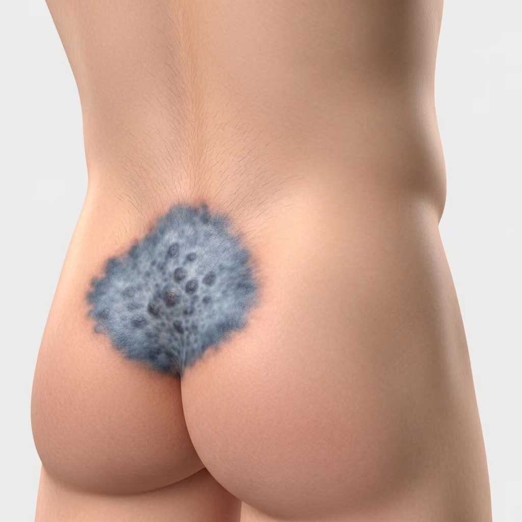
Lumbosacral dermal melanocytosis, commonly known as Mongolian spot, is a benign congenital birthmark characterized by blue-grey pigmentation on the skin, primarily affecting the lower back and buttock region of newborns.
What is lumbosacral dermal melanocytosis?
Lumbosacral dermal melanocytosis refers to flat, bluish-grey patches present at birth, most frequently located over the lumbosacral area, buttocks, and sometimes extending to the upper posterior thighs or flanks. These lesions result from melanocytes—pigment-producing cells—that become entrapped in the dermis during fetal development, failing to migrate fully to the epidermis. The Tyndall effect causes the deep dermal melanin to appear blue-grey on the surface, as shorter blue wavelengths scatter more superficially.
These birthmarks are macular (flat), non-palpable, and range from a few millimeters to over 10 cm in diameter. They do not blanch under pressure and show no accentuation under Wood’s lamp illumination. While typically solitary or few in number, extensive or multiple lesions can occur. The pigmentation is often darkest around 1 year of age and begins fading by age 3, with most resolving completely by age 4–5 years. Persistent cases, especially larger ones, may last into adulthood.
Who gets lumbosacral dermal melanocytosis (epidemiology)?
This condition is extremely common, occurring in over 90% of infants of East Asian, Polynesian, Indonesian, and Micronesian descent. Prevalence decreases in other groups: 46–100% in Hispanic newborns, 26–96% in Black infants, and 1–23% in Caucasian babies. It affects both sexes equally and shows no familial predisposition in most cases, though rare familial patterns exist.
Extra-sacral sites (shoulders, flanks, limbs) are more common in darker-skinned individuals, and persistent or atypical lesions warrant closer evaluation.
What causes lumbosacral dermal melanocytosis (aetiology)?
The primary cause is the arrested migration of melanocytes from the neural crest to the epidermal basal layer during embryogenesis (weeks 6–8 of gestation). These spindle-shaped melanocytes remain dispersed in the mid-to-deep dermis between collagen fibers.
No specific genetic mutations are identified in common cases, but extensive dermal melanocytosis associates with inborn errors of metabolism, such as lysosomal storage disorders (e.g., GM1 gangliosidosis type 1, mucopolysaccharidoses like Hurler’s and Hunter’s syndromes, Niemann-Pick disease). Phakomatosis pigmentovascularis links it to vascular malformations.
What are the clinical features of lumbosacral dermal melanocytosis?
Lesions appear at birth or within the first weeks as well-defined, homogenous blue-grey to blue-black macules or patches. Common locations include:
- Lumbosacral region (most frequent)
- Buttocks and gluteal cleft
- Flanks and upper thighs
- Rarely: shoulders, back, limbs, or face (if facial, consider nevus of Ota)
Skin texture remains normal without induration, scaling, or hair changes. Size varies from 5 mm to >10 cm; shapes are round, oval, or irregular. Color may deepen initially before fading.
In atypical cases:
- Multiple or extensive (>5% body surface)
- Persistent beyond age 5
- Midline spinal location (risk of spinal dysraphism)
- Accompanying hypertrichosis or lipoma (neural tube defect marker)

Diagnosis
Diagnosis is clinical based on characteristic appearance in a newborn, especially in high-prevalence ethnic groups. No routine testing is needed for typical cases.
Histology
Biopsy (rarely required) reveals elongated, dendritic melanocytes with abundant melanin in the deep dermis, oriented parallel to collagen bundles. No atypia or increased mitoses. Persistent lesions may resemble blue nevus with subcutaneous extension.
Differential diagnosis
| Condition | Key Distinguishing Features |
|---|---|
| Café-au-lait macule | Light brown, epidermal pigment; Wood’s lamp accentuates; persists |
| Blue nevus | Firmer, papular; persists into adulthood; histology shows nevus cells |
| Post-inflammatory hyperpigmentation | History of inflammation; not present at birth |
| Fixed drug eruption | Recurrent, targetoid; drug history |
| Vascular malformation/hemangioma | Blanching, compressible; ultrasound confirms |
| Linear/whorled nevoid hypermelanosis | Follows Blaschko lines; reticulated pattern |
| Child abuse (bruising) | Tender, evolves over days; multiple healing stages; extra-sacral |
Investigations
For typical solitary lumbosacral lesions: none required.
Indications for workup:
- Midline spinal lesion ± tuft of hair, lipoma, or dermal sinus (MRI spine for dysraphism)
- Extensive/persistent/multiple lesions (enzyme assays for lysosomal storage diseases)
- Atypical sites or rapid change (biopsy)
Management
Observation is standard; no treatment needed as lesions are benign and self-resolve. Educate parents on natural history to prevent unnecessary anxiety or misdiagnosis as bruising.
Persistent lesions (cosmetic concern): Q-switched ruby or Nd:YAG laser may lighten pigmentation, but recurrence is possible. Avoid in children unless severe.
Associated conditions: Refer to neurology/genetics/metabolism specialists as needed.
Complications
Typically none. Rare associations:
- Lysosomal storage disorders (GM1 gangliosidosis, mucolipidosis)
- Phakomatosis pigmentovascularis
- Occult spinal dysraphism (midline lesions)
Psychosocial: Misinterpretation as abuse in darker-skinned children.
Prevention
Not preventable, as it’s a developmental anomaly.
Prognosis
Excellent; >90% fade by school age. Persistent cases remain asymptomatic.
Frequently Asked Questions
Is lumbosacral dermal melanocytosis dangerous?
No, it is a harmless birthmark that usually disappears by age 4. Only atypical cases need evaluation.
Will my child’s Mongolian spot go away?
Yes, most fade completely by 3–5 years. Larger ones may persist but are benign.
Can Mongolian spots be mistaken for bruises?
Yes, especially in non-Caucasian infants. Document at birth to avoid child protection concerns.
Does it occur in all races?
Most common in Asian, Hispanic, Black infants (up to 90–100%); rare in Caucasians (1–23%).
Should I have it removed?
Not necessary; laser only for persistent cosmetic issues in adulthood.
Related topics
- Nevus of Ota
- Café-au-lait macules
- Blue nevus
- Phakomatosis pigmentovascularis
- Spinal dysraphism
References
- Dermal melanocytosis (previously known as Mongolian spot) — Dermatology Advisor. 2023. https://www.dermatologyadvisor.com/home/decision-support-in-medicine/dermatology/dermal-melanocytosis-previously-known-as-mongolian-spot/
- Lumbosacral dermal melanocytosis — DermNet NZ. 2003 (updated). https://dermnetnz.org/topics/lumbosacral-dermal-melanocytosis
- Dermal Melanocytosis (Mongolian Spots) — MD Searchlight. 2024. https://mdsearchlight.com/skin-problems-and-treatments/dermal-melanocytosis-mongolian-spots/
- Slate gray nevi (Mongolian blue spots) — Medical News Today. 2023-05-23. https://www.medicalnewstoday.com/articles/318853
- Dermal Melanocytosis — StatPearls, NCBI Bookshelf. 2023-08-08. https://www.ncbi.nlm.nih.gov/books/NBK557408/